
IBS Beyond Symptoms: What Faecal Proteases and Enteric Neuronal Activation May Be Telling Us
A translational Gut study suggests that stool-derived mediators activate enteric neurons differently in IBS-D and IBS-C, highlighting protease, immune, and biomarker pathways for future research.


When the Colonoscopy Is Normal but the Symptoms Clearly Are Not
Anyone who has spent enough years in a gastroenterology clinic knows this consultation.
The colonoscopy is normal. Routine blood tests offer little explanation. There is no obvious structural disease. Yet the patient sitting opposite you is describing urgency, abdominal pain, unpredictable bowel movements, and a quality of life that is anything but normal.
In another room, a patient with constipation-predominant IBS may have equally disabling symptoms but an almost opposite bowel pattern.
We classify both under irritable bowel syndrome. Clinically, that is useful. Biologically, it may be rather crude.
The paper “Faecal proteases and immune signatures drive subtype-specific enteric neuronal activation in IBS,” published Online First in Gut in August 2026, asks whether something measurable in the intestinal lumen helps explain why IBS-D and IBS-C behave differently.
The investigators studied whether faecal material from patients with IBS could directly activate enteric neurons and, importantly, whether the mechanism of that activation differed between diarrhoea-predominant and constipation-predominant disease.
This was a translational mechanistic study.
It was not a treatment trial.
It was not designed to validate a diagnostic test for routine practice.
And it certainly does not mean we should begin ordering stool protease panels on Monday morning.
What it does provide is an intriguing biological bridge between the contents of the intestinal lumen and altered enteric neuronal signalling.
For a disorder still defined largely by symptoms, that is worth examining carefully.
Why Look at the Enteric Nervous System?
The enteric nervous system is sometimes described as the gut's “second brain,” a phrase that has probably appeared on more patient-information leaflets than most of us care to count.
The physiology behind the cliché, however, is important.
Enteric neurons participate in the regulation of motility, secretion, vascular responses, sensory signalling, and communication between the mucosa and immune system.
Submucous plexus neurons are particularly interesting because they sit close to the interface between luminal contents and mucosal function.
IBS research has already implicated several biological processes, including altered epithelial permeability, visceral hypersensitivity, low-grade immune activity, microbial influences, and faecal protease activity.
What has been less clear is whether mediators present in stool can directly alter enteric neuronal activity—and whether this differs between recognised IBS subtypes.
That was the biological question behind this study.
If stool-derived material from IBS patients activates enteric neurons more strongly than material from healthy individuals, it would support the existence of a functional lumen–enteric nervous system pathway.
It would not solve IBS.
But it would move the discussion one step beyond describing symptoms.
What the Investigators Actually Did
According to the accessible abstract, investigators analysed faecal supernatants from:
21 patients with IBS-D
9 patients with IBS-C
18 healthy controls
Participants were recruited across centres in three countries.
The faecal supernatants were applied to submucous plexus neurons from guinea pig distal colon, and neuronal activation was measured using neuroimaging techniques.
The investigators also examined faecal proteolytic activity and performed proteomic profiling.
This design deserves some attention because it determines how far the findings can reasonably be taken.
The exposure was not a drug or a dietary intervention.
It was the soluble material present in stool from different clinical groups.
The researchers were essentially asking whether that luminal material could provoke a measurable neuronal response under controlled experimental conditions.
That is a useful mechanistic experiment.
But guinea pig enteric neurons exposed experimentally to human stool supernatant are not the same thing as a patient experiencing IBS symptoms in vivo.
The model allows us to study biological plausibility.
It does not reproduce the entire human disorder.
Both IBS Subtypes Activated Neurons—But Through Different Routes
One of the central findings was that faecal supernatants from both IBS-D and IBS-C patients produced significantly greater enteric neuronal activation than supernatants from healthy controls.
That observation alone is interesting.
It suggests that the stool of patients with IBS contains soluble mediators capable of influencing enteric neuronal behaviour.
The more important finding, however, was that the apparent mechanism differed between the two IBS subtypes.
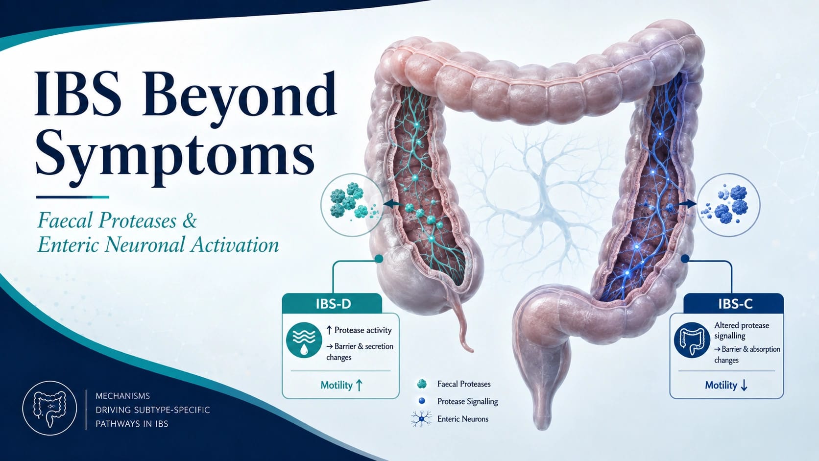
In IBS-D, neuronal activation was linked to:
serine proteases
cysteine proteases
protease-activated receptor-1, or PAR-1
In IBS-C, increased neuronal activation was also observed, but it appeared to occur independently of the same protease/PAR-1 pathway.
That distinction is arguably the most important contribution of the paper.
IBS-D and IBS-C may share a general phenomenon—enhanced luminal activation of enteric neurons—while reaching that endpoint through different molecular pathways.
For clinicians accustomed to seeing IBS represented as one large diagnostic category divided mainly by stool form, that is an intellectually useful shift.
The bowel habit may be the visible phenotype.
The biology underneath it may be considerably less uniform.
Proteases May Be Participants, Not Merely Passengers
Proteases are enzymes that cleave proteins, and the gastrointestinal lumen contains plenty of them.
Their sources may include pancreatic secretion, host tissues, inflammatory cells, microbial activity, and other luminal processes.
Faecal serine and cysteine protease activity has previously attracted attention in IBS because of possible associations with epithelial barrier dysfunction and visceral hypersensitivity.
This study extends that conversation toward the enteric nervous system.
In IBS-D, inhibition experiments implicated protease activity and PAR-1 in the neuronal response generated by faecal supernatants.
That is more informative than simply finding that protease concentrations differ between groups.
It suggests a functional pathway through which luminal proteolytic activity may influence neuronal signalling.
The distinction between association and mechanism, however, remains important.
The study supports a mechanistic role for proteases in an experimental system.
It does not establish that proteases are the sole—or even dominant—cause of symptoms in every patient with IBS-D.
It also does not demonstrate that inhibiting those proteases will improve pain, urgency, stool frequency, or quality of life.
That therapeutic leap remains to be tested.
A Subtle Immune Signal in IBS-D
Proteomic analysis added another layer.
The investigators reported significant differences in 47 proteins between IBS-D patients and healthy controls. Several of these involved immunoglobulin components.
The authors interpreted these findings as compatible with an immune-enriched or microinflammatory faecal signature.
This requires careful language.
IBS is not inflammatory bowel disease.
Nothing in this study suggests that IBS-D should suddenly be viewed as a mild form of ulcerative colitis or Crohn's disease.
The term microinflammation refers here to subtle immune-associated molecular signals—not macroscopic inflammation, ulceration, or the structural tissue injury we associate with IBD.
This may help explain an experience clinicians encounter frequently: patients can have substantial symptoms despite normal conventional structural testing.
“Normal colonoscopy” does not necessarily mean “nothing biological is happening.”
Nor, of course, does every detectable molecular difference automatically become clinically important.
The challenge is determining which signals genuinely contribute to symptoms and which simply travel alongside them.
Could These Signals Become Biomarkers?
The study also identified a combination involving amylases, trypsin-2, and an immunoglobulin protein that showed high diagnostic performance in distinguishing IBS-D from healthy controls.
For anyone who manages IBS regularly, the attraction is obvious.
IBS remains primarily a clinical diagnosis. A biological marker reflecting disease mechanism rather than nonspecific inflammation would be valuable.
A stool-based panel that helped identify a particular IBS-D phenotype could eventually support more objective disease stratification.
Eventually is the important word.
This study does not establish a clinically validated diagnostic assay.
The study population was small, particularly the IBS-C group.
Diagnostic performance derived from a discovery cohort can look impressive and then diminish considerably when tested in different populations.
A clinically useful biomarker would need to work not only against healthy volunteers but against patients with the conditions clinicians actually struggle to distinguish from IBS-D.
That includes inflammatory disease, infection, bile acid diarrhoea, malabsorption, medication-related diarrhoea, and other gastrointestinal disorders.
A biomarker that separates IBS-D from perfect health is scientifically interesting.
A biomarker that helps us manage the patient in front of us is a higher bar.
What This May Tell Us About IBS-D
For IBS-D, the study offers a reasonably coherent mechanistic model.
Faecal material contains proteolytic and immune-associated signals.
Those signals can activate enteric neurons experimentally.
Part of that neuronal activation appears to involve serine and cysteine proteases and PAR-1.
Biologically, one can see why this attracts attention.
Enhanced neuronal activation could potentially contribute to phenomena familiar in IBS-D—urgency, altered sensory processing, abnormal secretion, and symptom fluctuations despite an absence of structural disease.
But this remains a hypothesis supported by experimental data, not a clinical treatment pathway.
The study does not demonstrate that:
protease inhibition improves IBS-D symptoms
PAR-1 blockade is an effective therapy
all patients with IBS-D share the same protease signature
stool protease measurement can guide treatment selection
established IBS-D therapies should be replaced
The proper interpretation is that the study identifies a plausible mechanistic pathway worthy of therapeutic investigation.
That is already valuable.
There is no need to make it promise more.
IBS-C May Be the More Interesting Unfinished Story
IBS-C deserves equal attention.
Faecal material from IBS-C patients also produced greater neuronal activation than material from healthy controls.
But that activation did not appear to depend on the same protease/PAR-1 mechanism identified in IBS-D.
That finding is useful precisely because it leaves a question unanswered.
What is activating those neurons in IBS-C?
This study does not provide a definitive answer.
Other luminal mediators may be involved, but the source does not establish which ones.
It would therefore be premature to fill the gap with speculation about metabolites, bile acids, microbial products, or other candidate pathways.
What the paper does show is simpler and more defensible:
The neuronal activation observed in IBS-C appears biologically distinct from the protease-dependent pathway identified in IBS-D.
The smaller IBS-C sample—only nine patients in the accessible abstract—also means that this observation should be regarded as exploratory.
Still, it is an important warning against assuming that mechanisms discovered in diarrhoea-predominant IBS automatically apply to constipation-predominant disease.
Clinical labels sometimes share a surname while having rather different relatives underneath.
Why the Study Design Is Strong
Several aspects of the study strengthen its biological argument.
First, the investigators used human faecal material from clinically characterised IBS-D and IBS-C patients.
Second, they measured functional neuronal activation, rather than stopping at static molecular associations.
Third, they combined those functional experiments with measurements of protease activity and proteomic profiling.
Fourth, they analysed IBS-D and IBS-C separately rather than treating IBS as a single biological entity.
That integration is important.
Finding a different protein concentration in stool is interesting.
Showing that stool-derived material produces a neuronal response—and then investigating which molecular pathways contribute to that response—provides a more coherent mechanistic story.
That remains a laboratory story for now.
But it is a stronger one.
Where We Need to Be Cautious
There are several reasons not to move too quickly from these findings to clinical practice.
The neuronal model was not human
The experiments used guinea pig distal colon submucous plexus neurons.
This is an established way to study enteric neuronal responses, but it cannot reproduce the complete physiology of the human gastrointestinal tract.
The sample was small
The accessible abstract describes 21 IBS-D patients, nine IBS-C patients, and 18 healthy controls.
Those numbers may be sufficient for exploratory mechanistic work but are far from adequate for definitive clinical phenotyping or diagnostic validation.
Stool composition is influenced by many variables
Diet, medication, microbiome composition, transit time, host immune activity, and other environmental or biological factors can influence faecal proteins and proteolytic activity.
The study does not establish how stable these signatures are over time.
Neuronal activation is not the same as symptom causation
Demonstrating that a faecal supernatant activates neurons experimentally does not prove that the same pathway explains abdominal pain, urgency, stool frequency, bloating, or the entire clinical IBS phenotype.
The proposed biomarkers remain investigational
The reported protein combination is a research finding.
It is not ready for routine stool testing or clinical decision-making.
What Clinicians Should Not Conclude
This study does not show that IBS-D is simply a protease disorder.
It does not show that IBS-C has now been mechanistically explained.
It does not justify faecal protease testing in routine gastroenterology practice.
It does not establish PAR-1 inhibition as treatment.
And it does not make current dietary, pharmacological, behavioural, or other established IBS management approaches obsolete.
Those conclusions would travel substantially further than the evidence.
A more appropriate interpretation is that IBS subtypes appear capable of generating biologically distinct stool-derived signals that influence enteric neuronal activity.
For a condition historically defined largely by symptoms, that is meaningful progress.
It is not yet a prescription.
The Larger Question: Are We Moving Toward Biological IBS Subtypes?
Perhaps the most interesting implication of this work is not one particular protease or one potential stool biomarker.
It is the possibility that the familiar IBS-D and IBS-C labels may eventually sit on top of more specific biological phenotypes.
At present, treatment remains largely symptom-directed.
A future approach might classify patients according to combinations of luminal mediators, microbial function, immune activity, neural sensitivity, epithelial barrier characteristics, and clinical phenotype.
That possibility is attractive.
But gastroenterology has seen many promising biomarkers arrive early and leave quietly.
The next steps therefore matter enormously.
The proteomic findings need independent replication.
The neuronal findings need confirmation.
The proposed biomarkers need validation in larger and more diverse populations.
Researchers need to determine whether these molecular signatures correlate with clinically important outcomes such as:
abdominal pain
urgency
stool frequency
visceral hypersensitivity
symptom severity
treatment response
Longitudinal studies would also be useful.
Do these protease and immune signatures fluctuate when symptoms worsen?
Do they normalise when patients improve?
Or are they stable traits identifying biologically distinct subgroups?
Those questions will determine whether these findings become clinically useful or remain elegant mechanistic observations.
From Mechanism to Treatment Will Require Another Step
The study also opens an obvious therapeutic question.
If protease/PAR-1 signalling contributes meaningfully to IBS-D biology, could that pathway be targeted?
Possibly.
But mechanistic plausibility is not therapeutic efficacy.
Interventional studies would need to identify the correct patient population, demonstrate target engagement, and—most importantly—show improvement in outcomes that matter to patients.
A pathway can look beautiful on a laboratory diagram and still disappoint in the clinic.
Gastroenterology has provided us with enough examples to remain appropriately humble.
The same principle applies to biomarker development.
Before a faecal protein panel becomes useful, researchers will need to demonstrate that it adds something beyond careful clinical assessment and that it performs well against relevant competing diagnoses.
Only then does mechanistic elegance become clinical utility.
Clinical Takeaway
This Gut study provides experimental evidence that faecal mediators from patients with IBS can activate enteric neurons more strongly than faecal material from healthy controls.
More importantly, the mechanism appears to differ by subtype.
In IBS-D, neuronal activation was associated with serine and cysteine proteases and PAR-1, alongside an immune-enriched faecal proteomic signature.
IBS-C faecal supernatants also increased neuronal activation, but apparently through a different and currently undefined pathway.
For practicing gastroenterologists, this should not change tomorrow's treatment algorithm.
IBS remains a clinical diagnosis, and the proposed faecal biomarkers are not ready for routine testing.
The value of the study is more fundamental.
It strengthens the argument that IBS-D and IBS-C may represent biologically distinct disorders beneath their symptom-based labels—and that stool-derived signals can interact directly with the enteric nervous system.
That brings us closer to a mechanistic understanding of IBS.
Whether it eventually brings us better biomarkers or more targeted treatments will require much larger and more clinically oriented studies.
For now, the appropriate response is interest without overenthusiasm.
In IBS research, that is usually a sensible place to stand.
Five Key Clinical Takeaways
The study is mechanistic, not a clinical trial. Human faecal supernatants were tested on enteric neurons to investigate biological pathways underlying IBS subtypes.
Both IBS-D and IBS-C stool-derived mediators increased neuronal activation. This supports a potential lumen–enteric nervous system signalling pathway in IBS.
IBS-D showed a distinct protease-related mechanism. Serine and cysteine proteases and PAR-1 were implicated in neuronal activation, whereas IBS-C activation appeared independent of this pathway.
The faecal proteomic findings are promising but preliminary. A candidate protein combination showed diagnostic potential for IBS-D, but it is not a validated clinical test.
Nothing here justifies changing current IBS management. The study supports biological subtyping and future biomarker and therapeutic research, not immediate changes to diagnosis or treatment.

Source Reference
Ridžal L, Frieling T, Róka R, et al. Faecal proteases and immune signatures drive subtype-specific enteric neuronal activation in IBS. Gut. Online First, August 2026.

We are pioneers in clinical intelligence, dedicated to helping gastroenterologists harness the power of artificial intelligence to drive precision, efficiency, and patient growth.