
Machine Learning for Liver Fibrosis Prediction in MASLD: Promising Tool or Premature Clinical Leap?
Machine learning may improve fibrosis risk prediction in MASLD, but validation, calibration, and clinical workflow integration remain essential.


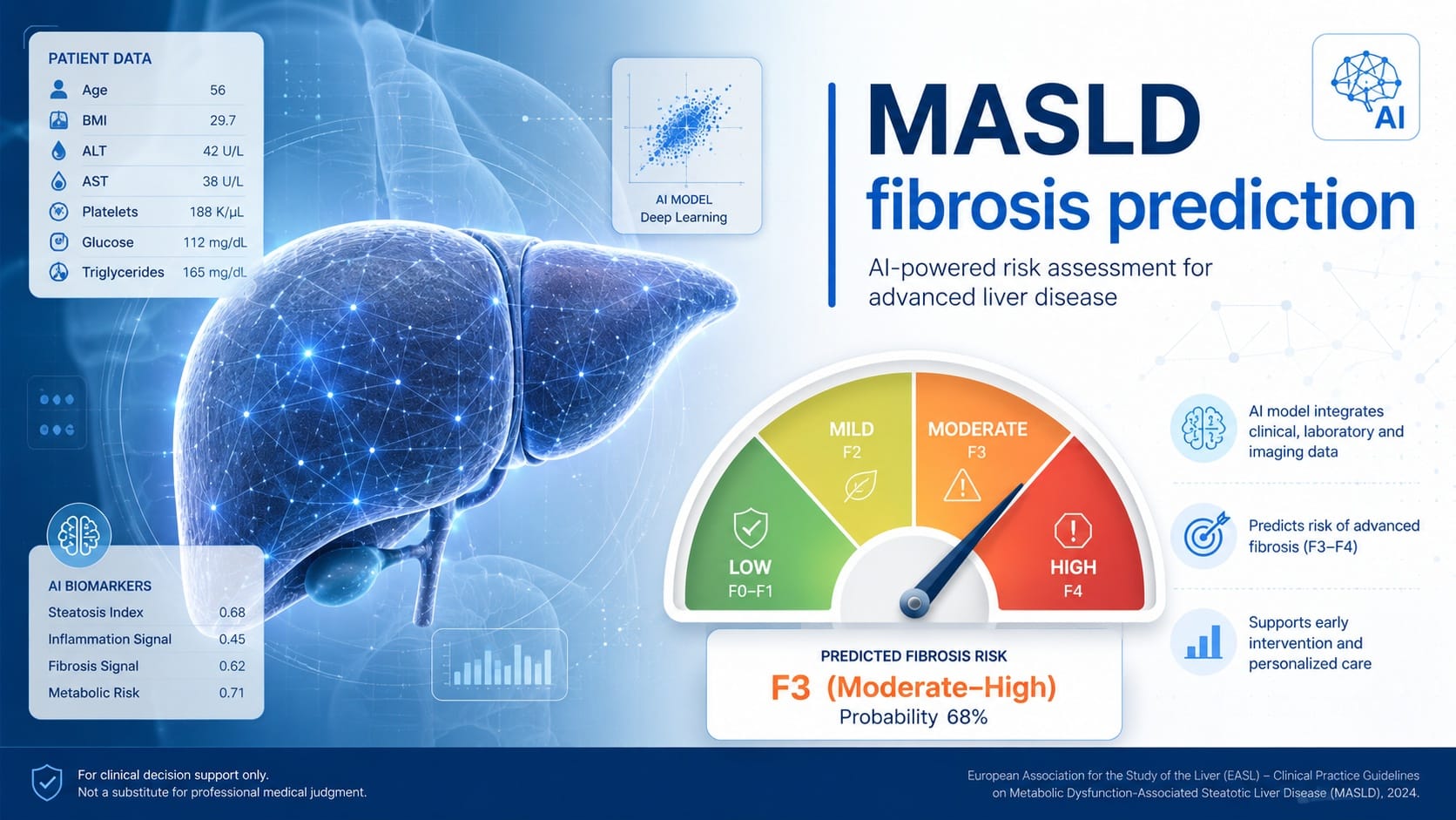
Can machine learning help gastroenterologists identify clinically important fibrosis in MASLD more accurately than the non-invasive tools already used in practice?
That is the central question raised by the conference paper “Machine Learning Prediction of Liver Fibrosis in Patients with Metabolic Dysfunction Associated Steatotic Liver Disease,” published in Digestive and Liver Disease, Volume 58, Supplement 1, February 2026, page S130, as abstract F-80. The work is authored by Salvatore Petta, Grazia Pennisi, Ciro Celsa, Sofiane Messaoudi, Emmanuel Tsochatzis, Elisabetta Bugianesi, Masato Yoneda, Ming-Hua Zheng, Hannes Hagström, Jérôme Boursier, José Luis Calleja, George Boon-Bee Goh, Wah-Kheong Chan, Rocio Gallego-Durán, Arun J. Sanyal, and collaborators. The DOI listed is 10.1016/j.dld.2026.01.204.
This is not a therapeutic trial and not a guideline. It is a machine-learning prediction-model development and validation study, reported as a conference paper/abstract. Its clinical focus is highly relevant: identifying liver fibrosis stage in patients with metabolic dysfunction-associated steatotic liver disease, or MASLD. The source explicitly frames the unmet need: accurate fibrosis staging is crucial; liver biopsy is invasive; and commonly used non-invasive tests such as FIB-4, liver stiffness measurement, and Agile 3+ may not always provide sufficient accuracy.
The clinical problem: fibrosis risk, not steatosis alone
For clinicians managing MASLD, the practical challenge is not simply detecting steatosis. The harder question is determining which patients have clinically meaningful fibrosis and therefore need closer assessment, follow-up, or specialist prioritization.
The abstract starts from this point. Liver biopsy remains a reference standard for staging fibrosis, but it is invasive and not suited to broad, repeated, routine risk stratification. In daily practice, clinicians rely on non-invasive tests. FIB-4 is simple and accessible. Liver stiffness measurement provides imaging-based assessment. Agile 3+ combines selected parameters to estimate advanced fibrosis risk. Yet each test leaves room for uncertainty, especially when patients fall into indeterminate zones or when results are discordant.
Machine learning is attractive in this setting because fibrosis risk may be influenced by multiple clinical and laboratory variables that interact in ways not easily captured by a fixed linear formula. The study investigated whether models trained on routinely collected clinical data could improve fibrosis assessment in biopsy-proven MASLD. This is the right type of question for AI in hepatology: not replacing clinicians, but potentially improving triage and staging where current tools are imperfect.
What the investigators studied
The investigators developed machine-learning models using a training cohort of biopsy-proven MASLD patients. The models incorporated 22 clinically relevant variables, including liver stiffness measurement. Internal validation used a 25% random test split across five MICE-imputed datasets, and external validation was performed in three independent cohorts from different centers.
Several model types were evaluated. These included FT-Transformer, TabNet with and without liver stiffness measurement, and two ordinal models: MLP-CORAL and CoralTabNet. The binary prediction tasks focused on detecting fibrosis thresholds of F≥3, F≥2, and F4. The authors also evaluated multiclass staging across F0 to F4.
This design matters clinically because it does not only ask whether a model can identify cirrhosis. It also asks whether machine learning can distinguish fibrosis thresholds that may influence monitoring intensity, referral decisions, and staging confidence. The use of biopsy-proven MASLD patients anchors the model against histological staging, while external validation across three cohorts is an important strength for assessing generalizability.
At the same time, the accessible source is an abstract. It does not provide all details needed for full methodological appraisal, such as exact sample size, inclusion and exclusion criteria, the complete list of variables, missing data proportions, model calibration, decision-curve analysis, subgroup performance, or how the three external cohorts differed. These gaps are important when considering clinical adoption.
Key findings: better performance, but not uniformly superior
For detection of F≥3 fibrosis, FT-Transformer and TabNet achieved AUCs of 0.860 and 0.855, respectively. The reported grey zones were small: 8.2% and 8.4%. These models outperformed FIB-4, which had an AUC of 0.756, and liver stiffness measurement alone, reported with an AUC of 0.837. However, they did not significantly outperform Agile 3+, which had an AUC of 0.847 with a reported p value of 0.12.
For F≥2 fibrosis, FT-Transformer and TabNet reached AUCs of 0.800 and 0.794. These exceeded FIB-4, reported at 0.713, and liver stiffness measurement, reported at 0.775. Again, the models did not meaningfully outperform Agile 3+, which had an AUC of 0.794 and a p value of 0.548.
For F4 fibrosis, FT-Transformer and TabNet achieved AUCs of 0.834 and 0.831, with the abstract stating that they outperformed conventional non-invasive tests. In multiclass staging from F0 to F4, MLP-CORAL showed the best agreement with biopsy, with a quadratic weighted kappa of 0.616. Models without liver stiffness measurement performed consistently worse across tasks. The authors report that findings were confirmed across all three external validation cohorts, with stable AUROC values and minimal grey zones.
These findings suggest that machine-learning models, especially FT-Transformer and TabNet, may improve discrimination compared with some established non-invasive tools. The most clinically relevant nuance is that superiority was not universal. For important thresholds such as F≥3 and F≥2, the models outperformed FIB-4 and liver stiffness measurement in the reported results, but did not significantly outperform Agile 3+. That distinction matters. It prevents overinterpreting the study as showing that machine learning is clearly better than all existing tools.
Why liver stiffness measurement still matters
One of the most clinically useful findings is that models without liver stiffness measurement performed worse across all tasks.
This has two implications. First, machine learning did not appear to eliminate the value of elastography-derived information. Instead, liver stiffness measurement remained an important component of model performance. Second, a model requiring liver stiffness measurement may be less immediately scalable in settings where elastography access is limited.
That does not make the model unhelpful. It simply clarifies the use case. A machine-learning tool that integrates liver stiffness measurement may be best positioned as an enhanced interpretation layer after elastography, not necessarily as a universal first-line screening test based only on routine blood work.
For busy hepatology and gastroenterology services, this distinction is practical. If the model requires liver stiffness measurement, it may improve staging confidence after a patient has already entered a liver assessment pathway. If future versions can perform well without liver stiffness measurement, they may be more useful in primary-care triage or large population-level MASLD screening. The abstract reports that models without liver stiffness measurement performed worse, so that broader use case remains less supported by this source.
Practical interpretation for clinicians
The reported results are encouraging, but they should be interpreted as prediction-model evidence, not clinical guidance.
A model with an AUC around 0.86 for F≥3 fibrosis is potentially useful, especially if it reduces the number of indeterminate results. The abstract’s report of small grey zones is clinically interesting because grey-zone results are a common limitation of non-invasive fibrosis algorithms. If reproduced in full manuscripts and prospective care pathways, reducing uncertainty could help clinicians make clearer decisions about further testing or referral.
However, clinicians should not conclude that this model is ready to replace biopsy in every ambiguous case. The authors conclude that machine-learning models integrating routine clinical variables perform better than commonly used non-invasive tests and may provide an efficient, widely applicable alternative to biopsy in routine clinical care. That is the study’s conclusion, but from a clinical implementation standpoint, several questions remain unanswered in the accessible source.
The abstract does not report whether use of the model improves patient outcomes. It does not show whether the model changes referral patterns, reduces unnecessary biopsy, prevents missed advanced fibrosis, or improves cost-effectiveness. It also does not provide workflow details: where the model would sit in the MASLD care pathway, which threshold should trigger elastography or hepatology referral, and how clinicians should manage discordant results between the model, FIB-4, liver stiffness measurement, and Agile 3+.
Therefore, the best interpretation is balanced: this study supports machine learning as a promising adjunct for fibrosis staging in MASLD, particularly when liver stiffness measurement is available, but it does not establish a new standard of care.
Association, prediction, and causation
This study should be understood as a prediction study. It does not test a causal intervention. The machine-learning models identify patterns associated with fibrosis stage as defined against biopsy and fibrosis thresholds. They do not prove that any individual variable causes fibrosis progression, nor do they show that using the model will improve liver-related outcomes.
That distinction is especially important in AI-based clinical research. A model may predict fibrosis accurately because it identifies statistical patterns in the dataset. Those patterns can be clinically useful, but they are not the same as biological causation. For example, if a variable contributes strongly to prediction, that does not mean modifying that variable will necessarily change fibrosis stage. Prediction can guide risk stratification; causation guides intervention.
For GastroAGI readers, the central message is that the model may help identify who is more likely to have advanced fibrosis, but it should not be used to infer mechanistic drivers of fibrosis unless supported by separate biological or interventional evidence.
Strengths of the evidence
Several strengths make this abstract worth attention.
First, the study uses biopsy-proven MASLD patients for model development. Histology-based staging provides a clinically meaningful reference point for fibrosis classification, even though biopsy itself has known practical limitations.
Second, the investigators evaluated multiple clinically relevant fibrosis thresholds: F≥2, F≥3, F4, and multiclass F0–F4 staging. This is more informative than focusing only on cirrhosis or only on advanced fibrosis.
Third, the models were internally validated with a held-out 25% test split across five MICE-imputed datasets, suggesting attention to missing data and internal performance assessment.
Fourth, external validation was performed in three independent cohorts from different centers. External validation is essential for AI models because performance in a development cohort often overestimates real-world utility.
Finally, the comparison with established non-invasive tests provides clinical context. A model’s performance is meaningful only if compared with what clinicians already use. The abstract reports comparisons with FIB-4, liver stiffness measurement, and Agile 3+.
Limitations and unanswered questions
The most important limitation is that the available source is a conference abstract, not a full peer-reviewed manuscript with complete methods and supplementary details. It provides promising performance metrics but not the level of information needed for full clinical appraisal.
Several questions remain open. What was the sample size in the training and external validation cohorts? What were the inclusion criteria? Were patients recruited from tertiary hepatology clinics, metabolic clinics, or broader clinical settings? What was the prevalence of F≥2, F≥3, and F4 disease? How did performance vary across age, sex, diabetes status, body mass index, alcohol exposure, ethnicity, and geography? Were calibration plots reported? Were decision-curve analyses performed? How were clinically actionable thresholds selected?
These details matter because MASLD populations are heterogeneous. A model performing well in biopsy-enriched cohorts may behave differently in primary care or population screening, where advanced fibrosis prevalence is lower. Similarly, a model requiring liver stiffness measurement may be less applicable where elastography is unavailable or technically unreliable.
The source also does not establish prospective clinical utility. Before implementation, a model should ideally be tested in real clinical workflows to determine whether it improves appropriate referrals, reduces unnecessary testing, shortens diagnostic pathways, or identifies advanced fibrosis earlier without increasing harm.
What clinicians should and should not conclude
Clinicians can reasonably conclude that machine-learning models integrating routine clinical variables and liver stiffness measurement showed promising discrimination for fibrosis staging in biopsy-proven MASLD cohorts. They can also conclude that FT-Transformer and TabNet performed better than FIB-4 and liver stiffness measurement alone for selected fibrosis thresholds in the reported results, but not significantly better than Agile 3+ for F≥3 and F≥2.
Clinicians should not conclude that machine learning has replaced established MASLD fibrosis pathways. They should not use these findings to avoid biopsy when biopsy is otherwise clinically indicated. They should not assume the model is validated for every healthcare setting, every ethnicity, every disease prevalence, or every elastography platform. They should also not interpret the model as identifying causal drivers of fibrosis progression.
Most importantly, clinicians should resist the common AI trap: equating improved AUC with immediate clinical adoption. AUC is useful, but clinical implementation requires interpretability, calibration, reproducibility, workflow integration, safety monitoring, and clear action thresholds.
Future research and implementation priorities
The next step should be publication of full methods and prospective validation. Future studies should clarify the exact variables used, model calibration, subgroup performance, decision thresholds, and implementation strategy.
Prospective studies should ask whether model-guided care changes decisions. Does it reduce the indeterminate group? Does it improve selection for elastography, hepatology referral, biopsy, or surveillance? Does it perform well in lower-prevalence populations? Can it be embedded into electronic health records without adding clinician burden? Can clinicians understand and trust the output?
Another priority is comparison against complete care pathways, not isolated tests. In practice, clinicians do not use FIB-4 in isolation; they combine non-invasive tests with metabolic risk profile, imaging, longitudinal trends, and clinical judgment. Machine learning should be judged against this real-world standard.
Clinical Takeaway
This Digestive and Liver Disease conference paper suggests that machine-learning models, particularly FT-Transformer and TabNet, may improve fibrosis prediction in biopsy-proven MASLD when routine clinical variables and liver stiffness measurement are integrated. The findings are clinically relevant because fibrosis staging remains central to MASLD management, and current non-invasive tests leave room for uncertainty.
However, the evidence remains abstract-level prediction-model research. It is promising, not practice-changing. The study supports further validation and clinical implementation research, but it does not yet justify replacing established non-invasive pathways, biopsy when clinically needed, or clinician judgment.
Five key clinical takeaways
Verified source: This is a Digestive and Liver Disease conference paper/abstract, not a CGH full article, published in February 2026.
Study design: Machine-learning prediction-model development with internal validation and external validation in three independent cohorts.
Population: Biopsy-proven MASLD patients; exact sample size was not available in the accessible abstract.
Main finding: FT-Transformer and TabNet showed promising AUCs for F≥3, F≥2, and F4 fibrosis, outperforming FIB-4 and liver stiffness measurement in reported comparisons, but not Agile 3+ for F≥3 or F≥2.
Clinical interpretation: This is promising AI-based risk stratification evidence, but it remains hypothesis-generating and not yet practice-changing.
Source reference and link
Petta S, Pennisi G, Celsa C, et al. Machine Learning Prediction of Liver Fibrosis in Patients with Metabolic Dysfunction Associated Steatotic Liver Disease. Digestive and Liver Disease. 2026;58(Suppl 1):S130. DOI: 10.1016/j.dld.2026.01.204.
References

We are pioneers in clinical intelligence, dedicated to helping gastroenterologists harness the power of artificial intelligence to drive precision, efficiency, and patient growth.