
Find the Feeder: What Precision EUS Targeting May Change in Gastric Variceal Therapy
A 2026 Gastrointestinal Endoscopy commentary and supporting comparative study examine whether EUS-guided feeder-vessel targeting can reduce coil, glue, and reintervention requirements in selected gastric varices.


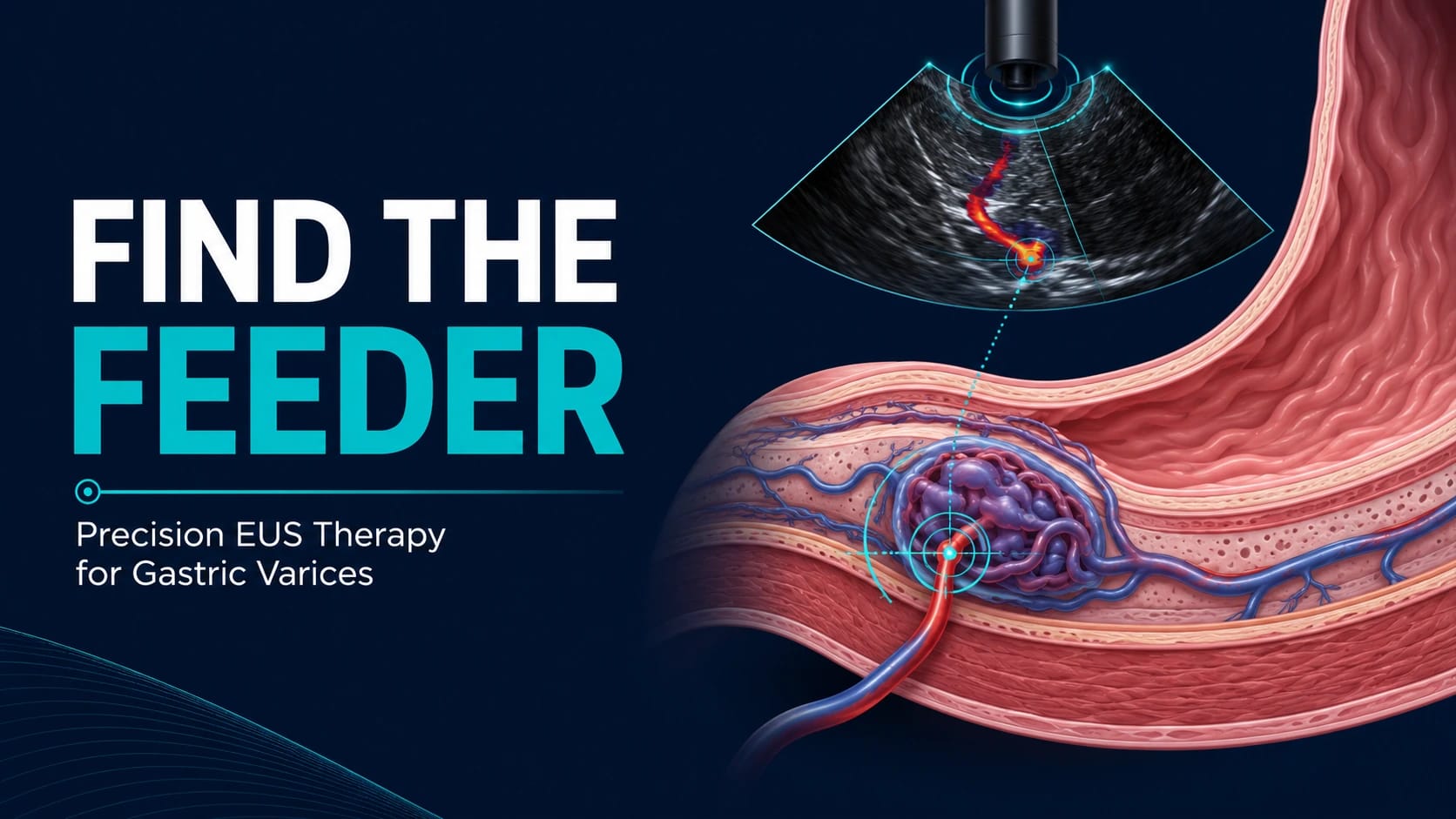
The Varix We See May Not Be the Best Target
When a gastric varix appears on endoscopy, the instinctive therapeutic target is the varix itself.
That makes perfect sense. It is visible, often impressive, and in the wrong clinical moment capable of producing a bleed nobody in the room will forget quickly.
EUS, however, changes the perspective.
Instead of showing us only the submucosal variceal complex, it can reveal the vascular architecture feeding it—collateral channels, inflow vessels, Doppler flow, and the relationship between the varix and surrounding vessels.
The recent Gastrointestinal Endoscopy commentary “Precision gastric variceal therapy: find the feeder,” by Cameron Thompson, published online on August 5, 2026, focuses on this idea.
The accompanying clinical question is straightforward:
If we can clearly identify the vessel feeding the gastric varix, should we treat the feeder rather than the visible variceal complex?
The commentary itself is not a new therapeutic trial. The comparative data behind the concept come from Dhar and colleagues, who examined EUS-guided coil-and-glue embolization directed either at the feeder vessel or at the submucosal variceal complex.
That distinction matters.
The commentary offers interpretation.
The supporting study provides pragmatic comparative data.
Neither should be read as definitive randomized evidence establishing feeder targeting as the universal standard for gastric varices.
What the Comparative Study Actually Asked
Dhar and colleagues evaluated patients undergoing EUS-guided coil-and-glue therapy in whom a discernible feeder vessel could be identified.
That qualification is central to the entire study.
The investigators compared two anatomical strategies:
EUS-guided treatment directed at the feeder vessel
EUS-guided treatment directed at the submucosal variceal complex
The primary outcome was reintervention rate.
Secondary outcomes included:
coil use
glue use
gastric variceal obliteration
number of EUS sessions
bleeding outcomes at 6 months
This was a pragmatic comparative analysis rather than a blinded randomized trial.
The question was therefore not whether EUS-guided coil-and-glue therapy works in gastric varices generally.
It was more technical:
Does choosing a different vascular target make the procedure more efficient?
For therapeutic endoscopists, that is a very practical question.
The Anatomy Behind the Idea
A gastric varix is not an isolated balloon sitting beneath the mucosa.
It is part of a collateral vascular circuit.
Conventional endoscopy shows us its luminal expression.
EUS allows us to see something closer to the plumbing.
The “find the feeder” strategy takes advantage of that additional information.
Rather than filling or embolizing the visible submucosal complex, the operator identifies the inflow vessel and attempts to interrupt the circulation supplying the varix.
The logic is attractive.
If one can control the vessel supplying the varix, perhaps fewer coils and less glue are required to achieve obliteration.
Anyone who has performed technically difficult vascular EUS will also appreciate the other side of the argument: anatomy is not always kind enough to present the feeder in a clean textbook plane.
The strategy only works when the vessel is clearly identified and safely accessible.
Who Was Studied?
The comparative analysis included 135 patients.
Of these:
32 patients underwent feeder-vessel targeting
103 patients underwent targeting of the submucosal variceal complex
GOV2 was the most common varix type, reported in 81 patients, representing 60% of the cohort.
Arakawa type 1 anatomy was reported in 97 patients, or 71.85%.
Both groups received EUS-guided coil-and-glue treatment.
The difference lay in where the operator delivered it.
That is worth emphasizing because the study is not comparing EUS-guided therapy with conventional glue injection, radiologic intervention, TIPS, or another portal hypertension strategy.
It compares two EUS-guided anatomical targets within a selected population.
What Happened When the Feeder Was Targeted?
The feeder-vessel approach was associated with lower use of both coils and glue.
Across the 6-month period, mean overall coil use was:
1.94 ± 0.91 in the feeder-vessel group versus 2.79 ± 1.76 in the submucosal variceal-complex group.
Mean overall glue use was:
1.88 ± 0.64 mL versus 2.83 ± 1.70 mL, respectively.
The study also reported a lower 6-month reintervention rate:
3.1% with feeder-vessel targeting versus 17.5% when the submucosal variceal complex was targeted.
Fewer EUS sessions were also required to achieve complete gastric variceal obliteration in the feeder-targeting group.
Taken together, these results support the possibility that a more anatomically precise target may achieve treatment goals with less material and fewer repeat procedures.
That is clinically interesting.
It is not yet the same thing as proving superiority in every patient.
Why Using Less Coil and Glue May Matter
At first glance, reduced coil and glue use might sound like a procedural detail best left to the supply room.
It deserves more attention than that.
EUS-guided gastric variceal therapy can be technically demanding and resource intensive.
Reducing material requirements could plausibly:
simplify the procedure
lower consumable costs
reduce the amount of embolic material deployed
decrease the need for additional treatment sessions
The investigators suggested that this might translate into a more cost-effective approach.
The word “might” is doing appropriate work here.
The accessible study information does not describe a formal economic analysis.
Therefore, lower material use should not be converted into a claim of proven cost-effectiveness.
Similarly, less glue exposure might sound intuitively attractive from a safety perspective, but this study does not establish that feeder targeting produces fewer embolic complications.
Those remain plausible hypotheses rather than demonstrated outcomes.
The More Interesting Shift Is From Appearance to Hemodynamics
The larger lesson may not be the exact number of coils used.
It is the change in therapeutic thinking.
Traditional endoscopy is largely lesion directed.
We identify what looks abnormal and treat it.
EUS adds another layer: flow-directed intervention.
In gastric varices, this means asking not only where the varix is located, but:
Where is the inflow?
How is the collateral network organized?
Which vessel can be safely accessed?
Can Doppler confirm the relevant flow before and after embolization?
This approach fits a broader evolution in advanced endoscopy.
We increasingly treat anatomy not simply as a static structure, but as a functional map.
The Dhar study supports that idea by suggesting that the choice of vascular target may influence procedural efficiency.
It should still be viewed as a signal requiring prospective validation.
What This Means for the Advanced Endoscopist
For clinicians already performing EUS-guided gastric variceal therapy, the study raises several useful questions before puncture.
Can the feeder vessel be identified confidently?
Is its course technically accessible?
Is the vessel large and stable enough for safe targeting?
Does the anatomy allow coil deployment without creating avoidable risk?
Is Doppler assessment adequate?
And perhaps most importantly, does the operator have sufficient experience to choose a feeder-targeted approach rather than simply pursue it because the concept is appealing?
The study only supports feeder targeting when the feeder vessel is feasible and discernible on EUS.
That should not be treated as an incidental footnote.
It is the clinical boundary around the entire strategy.
A poorly visualized feeder does not become a good target simply because a paper has made feeder targeting interesting.
Precision endoscopy is still supposed to be precise.
When the Conventional Variceal-Complex Approach May Still Be Reasonable
There will be situations in which the submucosal variceal complex remains the more practical target.
Examples may include:
unclear feeding anatomy
unstable scope position
poor visualization
technically unsafe feeder access
limited local experience with vascular EUS interventions
The study does not demonstrate that conventional targeting has become obsolete.
Nor does it show that feeder-targeting failure should lead to increasingly aggressive attempts to reach a vessel at the expense of procedural safety.
A new technical concept should sharpen judgment, not override it.
What Hepatologists Should Take From the Study
The implications extend beyond the endoscopy unit.
Gastric variceal bleeding sits at the intersection of advanced endoscopy, portal hypertension, interventional radiology, and liver disease management.
A patient with gastric varices is not simply carrying an endoscopic lesion.
The broader questions remain:
What is the underlying portal hypertensive anatomy?
What is the severity of liver disease?
Are radiologic interventions appropriate?
Is portal decompression required?
What is the likelihood of rebleeding?
What expertise is available locally?
The study does not answer those questions or replace established portal hypertension management.
Its contribution is narrower.
In selected patients already undergoing EUS-guided coil-and-glue treatment, targeting the feeder vessel may improve procedural efficiency when the vascular anatomy is clearly defined.
That makes the technique potentially valuable within a multidisciplinary gastric variceal programme rather than a standalone solution to portal hypertension.
The Evidence Is Encouraging, but the Groups Were Unequal
The results favouring feeder targeting are clinically appealing.
The design, however, should temper certainty.
Only 32 patients underwent feeder-vessel targeting compared with 103 in the submucosal variceal-complex group.
Because this was not described as a randomized trial in the accessible study information, treatment selection may have been influenced by anatomy, operator judgment, or technical feasibility.
That creates an important possibility:
Patients chosen for feeder targeting may have had anatomy that was more suitable for successful targeted embolization in the first place.
In other words, some of the apparent benefit could reflect patient selection rather than the target alone.
The data remain useful.
They simply need to be read with the correct level of confidence.
Six Months Is Useful—But Not the Whole Story
The reported outcomes extend to 6 months.
That is adequate to assess early reintervention and obliteration.
It does not answer every clinically relevant question.
We still need longer-term data on:
recurrent variceal flow
delayed rebleeding
repeated interventions
long-term obliteration
patient-centred outcomes
adverse events
interaction with subsequent portal hypertension therapies
Gastric varices belong to a chronic hemodynamic disease process.
A technically successful 6-month result does not necessarily tell us what happens several years later.
Expertise May Be the Largest Barrier to Generalisation
Feeder-vessel targeting is inherently operator dependent.
The endoscopist must identify the relevant vascular anatomy, interpret Doppler findings, choose the safest access point, deploy coils appropriately, administer glue, and confirm treatment response.
This level of EUS-guided vascular intervention is not uniformly available.
A technique that performs well in expert hands may be much less reproducible in lower-volume centres.
That does not weaken the concept.
It does mean that any move toward widespread adoption would need to address training, procedural standards, equipment, case selection, and centre experience.
The success of precision therapy often depends on precision infrastructure.
What Clinicians Should Not Conclude
This study does not prove that feeder-vessel targeting is superior for every gastric varix.
It does not show that feeder targeting eliminates bleeding risk.
It does not establish a universal EUS technique for all centres.
It does not demonstrate formal cost-effectiveness.
It does not establish superior long-term outcomes beyond the reported follow-up.
And it certainly does not make other portal hypertension interventions irrelevant.
The most defensible conclusion is narrower:
In patients with gastric varices in whom a feeder vessel could be clearly delineated on EUS, feeder-vessel targeting was associated with lower coil use, lower glue use, fewer EUS sessions, and a lower 6-month reintervention rate than targeting the submucosal variceal complex.
That is a strong procedural signal.
It remains a signal.
What Should Come Next?
The obvious next step is prospective validation.
A multicentre randomized or carefully designed prospective study could compare feeder-vessel and variceal-complex targeting using:
standardized definitions of feeder anatomy
predefined technical criteria
consistent coil-and-glue protocols
procedural adverse events
reintervention
rebleeding
long-term obliteration
costs
patient-centred outcomes
Future work should also determine how often a feeder vessel can actually be identified in routine practice.
That matters.
A strategy may perform extremely well in the subgroup in whom it is technically possible while being applicable to only a fraction of all patients.
Knowing that denominator will be essential before feeder targeting can be incorporated into a broader treatment algorithm.
Clinical Takeaway
The phrase “find the feeder” captures an appealing evolution in gastric variceal therapy.
Rather than treating only the visible variceal complex, EUS allows the endoscopist to map the vascular inflow sustaining it and, when the anatomy is favourable, target that inflow directly.
In the supporting pragmatic comparative analysis, feeder-vessel targeting was associated with less coil and glue use, fewer treatment sessions, and a lower 6-month reintervention rate than targeting the submucosal variceal complex.
Those findings deserve attention.
They do not yet justify a universal change in practice.
The feeder group was smaller, treatment allocation was not established as randomized in the accessible data, follow-up was limited to 6 months, and successful implementation depends heavily on EUS expertise and vascular anatomy.
For now, the message is best framed as precision without overreach.
If the feeder is clearly visible, safely accessible, and the operator has the appropriate expertise, it may be a more efficient therapeutic target.
If it is not, forcing the concept onto unsuitable anatomy would miss the point entirely.
In gastric variceal therapy, the future may indeed be increasingly hemodynamic.
But the first rule remains the same as it has always been in advanced endoscopy:
Understand the anatomy before you treat it.
Five Key Clinical Takeaways
The “find the feeder” publication is an expert commentary, not a new randomized trial. The supporting evidence comes from a pragmatic comparative analysis of EUS-guided coil-and-glue therapy.
The anatomical target mattered in the reported cohort. Feeder-vessel targeting was associated with lower overall coil and glue use than targeting the submucosal variceal complex.
Reintervention was lower at 6 months. The feeder group had a reported reintervention rate of 3.1% versus 17.5%, although the study design limits causal certainty.
The approach is selective, not universal. Feeder-vessel targeting is relevant only when the feeder can be clearly identified and safely accessed on EUS.
This does not replace broader portal hypertension management. Gastric variceal therapy should remain integrated with hepatology, radiology, liver disease severity, and local expertise.

Source References
Thompson C. Precision gastric variceal therapy: find the feeder. Gastrointestinal Endoscopy. Published online August 5, 2026. DOI: 10.1016/j.gie.2026.06.045.
Dhar J, Thakur R, Gupta P, Samanta J, et al. Endoscopic ultrasound-guided coil and glue embolization of gastric varices targeting feeder vessel versus submucosal variceal complex: A pragmatic comparative analysis (with videos). Gastrointestinal Endoscopy. 2026. DOI: 10.1016/j.gie.2026.05.011.
References
- References Thompson C. Precision gastric variceal therapy: find the feeder. Gastrointestinal Endoscopy. Published online August 5, 2026
- J, et al. Endoscopic ultrasound-guided coil and glue embolization of gastric varices targeting feeder vessel versus submucosal variceal complex: A pragmatic comparative analysis (with videos). Gastrointestinal Endoscopy. 2026

We are pioneers in clinical intelligence, dedicated to helping gastroenterologists harness the power of artificial intelligence to drive precision, efficiency, and patient growth.