
Risk-Based Pathology Reporting After ESD for Early GI Cancer: Why Standardisation Matters
International Gut consensus standards aim to standardise pathology reporting after ESD for early GI cancer and support risk assessment.


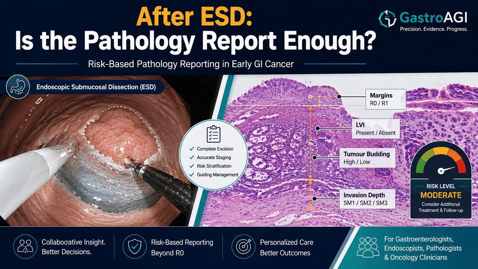
After endoscopic submucosal dissection for early gastrointestinal cancer, the endoscopy report tells only part of the story. The resection may appear technically successful, the lesion may have been removed en bloc, and the patient may leave the unit believing that cancer treatment is complete. But the next major clinical decision often depends on the pathology report: was the resection curative, or does the patient need further treatment?
That decision is rarely based on one histological feature alone. It depends on a risk-based synthesis of invasion depth, invasion breadth, margin status, lymphovascular invasion, tumour budding, differentiation, histological subtype, perineural invasion, specimen handling, and the reliability of the measurements reported. When these elements are reported inconsistently, multidisciplinary teams may struggle to determine whether surveillance is appropriate or whether additional therapy should be considered.
A new international consensus article in Gut, titled “Risk-based pathology reporting after endoscopic submucosal dissection for early gastrointestinal cancer: international consensus standards,” directly addresses this problem. It was published online ahead of print on 10 July 2026 and carries the DOI 10.1136/gutjnl-2025-337567. The work was led by Kareem Khalaf and colleagues and developed through an international modified Delphi consensus process.
A pathology report that determines the next clinical step
Endoscopic submucosal dissection, or ESD, enables en bloc resection of early gastrointestinal cancers and provides specimens that can be examined for detailed pathological risk assessment. The clinical strength of ESD is not only that it can remove selected lesions endoscopically, but that it preserves specimen architecture in a way that allows pathologists to assess features linked to residual disease risk, lymph node metastasis risk, and the need for additional treatment.
The problem identified by the consensus group is variability. The abstract specifically notes that reporting remains variable for key parameters that determine curative resection and the need for additional treatment. These include submucosal invasion depth and breadth, margin status, lymphovascular invasion, tumour budding, differentiation, and the use of ancillary stains.
For clinicians, this variability is not a technical inconvenience. It is a clinical problem. If one report describes invasion depth imprecisely, another uses different margin terminology, and a third omits tumour budding or lymphovascular invasion assessment, decision-making becomes less reliable. The same patient could be discussed differently depending on local pathology conventions rather than tumour biology.
This consensus article is therefore clinically relevant because it focuses on the interface between endoscopy, pathology, surgery, oncology, and surveillance planning. It is not a trial of ESD technique. It is not a drug study. It is a consensus standards paper designed to make post-ESD pathology reporting more reproducible and clinically meaningful.
What the consensus process investigated
The objective was to develop practical international standards for the pathology assessment and reporting of invasive carcinoma in ESD specimens. The study design was an international modified Delphi consensus process involving 42 experts from 15 countries. The panel included 28 gastrointestinal pathologists and 14 therapeutic endoscopists, which is important because the reporting standards were developed across the two specialties most directly involved in post-ESD interpretation and decision-making.
The consensus statements addressed several domains: measurement of invasion, margin assessment, staining, specimen handling, prognostic histological features, and clinically relevant reporting. The process resulted in 56 recommendations reaching consensus across seven domains.
The population here should be understood correctly. This was not a patient cohort study. There was no intervention group, no control group, and no patient-level clinical outcome analysis reported in the accessible abstract. The “population” for the consensus process was the international expert panel. The clinical material under consideration was ESD specimens containing invasive carcinoma from early gastrointestinal cancer. The “outcome” was consensus on practical pathology reporting standards, not survival, recurrence, lymph node metastasis, or cost-effectiveness.
That distinction matters. These standards may influence clinical practice by improving reporting consistency, but the abstract does not show that implementing them improves patient outcomes. It provides consensus-based reporting criteria intended to support clinical decision-making and future validation of risk models.
Measuring submucosal invasion: precision over habit
One of the most clinically important areas addressed is submucosal invasion. In early gastrointestinal cancers treated by ESD, the depth of invasion into the submucosa is a key element in risk assessment. The consensus panel recommends using the Sm1–Sm3 subclassification only when the muscularis propria is present. If the muscularis propria is not present, the depth of submucosal invasion should be reported in micrometres, rounded to the nearest 100 µm.
This recommendation is practical because the anatomical landmark used for subclassification may not always be present in an ESD specimen. If a classification system depends on a structure that is absent, reporting can become inconsistent or misleading. Reporting invasion depth in micrometres provides a more explicit measurement and reduces ambiguity.
The panel also recommends reporting submucosal invasion breadth in millimetres as an adjunct metric for future validation. This is a subtle but forward-looking point. The consensus group is not presenting breadth as a fully validated standalone decision rule in the accessible abstract. Instead, it identifies it as a metric that should be reported so future datasets can test its prognostic value more consistently.
For clinicians, the implication is clear: pathology reporting should not only describe that invasion is present, but define how it was measured and express it in reproducible terms. That does not automatically determine management by itself, but it improves the reliability of multidisciplinary interpretation.
Margin status: standard terminology for a high-stakes variable
Margin assessment is another central domain. The panel recommends that margin positivity should be defined as direct tumour contact with the inked surface. This definition is supported by standardised specimen pinning, inking, complete embedding, and parallel sectioning.
This matters because “positive,” “close,” “involved,” and “uncertain” margins can be used differently across institutions. In a post-ESD setting, margin status may influence whether a lesion is considered completely resected and whether additional treatment, repeat endoscopic therapy, surgery, or close surveillance is discussed. A precise definition helps reduce interpretive drift.
The consensus also links the margin definition to specimen handling. This is important because the quality of margin interpretation depends on what happens before the slide is read. Pinning, inking, embedding, and sectioning are not clerical details; they determine whether the pathologist can confidently assess the relationship between tumour and resection surface.
For endoscopists, this reinforces the need for close communication with pathology teams. A high-quality ESD specimen can still produce a less useful report if handling and orientation are inconsistent. Conversely, standardised pathology workflows can enhance the clinical value of an en bloc resection.
Ancillary stains: selective use, not automatic escalation
The consensus standards retain H&E as the baseline stain. Selective immunohistochemistry or elastic stains are recommended for situations such as equivocal lymphovascular invasion, distorted architecture, or difficult margin interpretation.
This is a balanced approach. It avoids implying that every ESD specimen requires extensive ancillary staining. At the same time, it recognises that certain clinically important features may be difficult to assess on routine staining alone.
Lymphovascular invasion is particularly relevant because it can influence risk stratification after endoscopic resection. The consensus does not state that ancillary stains should replace conventional histology. Rather, it supports selective use when interpretation is uncertain.
This distinction is important for clinicians reviewing reports. The absence of ancillary stains does not necessarily mean inadequate reporting if the case is straightforward. But when lymphovascular invasion or margins are equivocal, selective stains may improve interpretive confidence.
Tumour budding and composite risk: individual features need context
The panel recommends that tumour budding should be reported according to International Tumour Budding Consensus Conference criteria. It also states that differentiation, histological subtype, lymphovascular invasion, perineural invasion, and margin status should be integrated into composite risk assessment.
This is perhaps the core conceptual shift: pathology after ESD should not be a disconnected list of microscopic observations. It should support clinically relevant risk assessment.
Composite risk does not mean inventing a new score without validation. It means recognising that individual features interact in clinical decision-making. For example, a report that separately lists differentiation, invasion depth, lymphovascular invasion, and margins may be complete in a descriptive sense, but the clinician still needs to understand whether the combined profile suggests low-risk or higher-risk pathology.
The consensus standards aim to make these elements “synoptic-ready,” meaning suitable for structured reporting formats. Synoptic reporting is especially useful in multidisciplinary care because it reduces omission, improves clarity, and allows data aggregation for research and quality improvement.
Why this matters for multidisciplinary teams
The conclusion of the article states that these standards provide immediately implementable, synoptic-ready pathology reporting criteria after ESD. The authors also state that standardising measurement landmarks, margin terminology, ancillary stain use, and reporting of adverse histological features aims to reduce interinstitutional variability, improve multidisciplinary decision-making, and support future validation of risk models in early gastrointestinal cancer.
That conclusion is highly relevant to tumor boards and post-resection pathways. After ESD, the decision is often not binary. Some patients clearly meet criteria for endoscopic cure. Others clearly require further treatment. But many cases sit in the difficult middle: a close or uncertain margin, borderline invasion depth, equivocal lymphovascular invasion, or histological features that raise concern without providing a single definitive answer.
Standardised reporting does not eliminate clinical judgment. It improves the substrate on which clinical judgment is based.
For gastroenterologists and hepatologists involved in upper GI, colorectal, and early cancer pathways, the most practical value may be in reducing ambiguity at the moment when endoscopic therapy transitions into longitudinal cancer-risk management. For fellows and trainees, the paper also provides a useful reminder that pathology reports are not passive documents. They are active clinical tools.
What clinicians should conclude
Clinicians can conclude that an international expert panel reached consensus on 56 recommendations for pathology assessment and reporting after ESD for early gastrointestinal cancer. The standards address invasion measurement, margin definition, staining, specimen handling, histological risk features, and clinically relevant reporting.
They can also conclude that the paper supports more structured, risk-based, synoptic-ready reporting after ESD. This is especially relevant where endoscopic resection is being used for early invasive carcinoma and post-resection decisions depend heavily on histological risk features.
Clinicians should not conclude that this article proves improved survival, reduced recurrence, or reduced need for surgery. The accessible source does not report patient outcomes after implementation of these standards. It also does not validate a new risk model. Instead, it provides consensus criteria intended to improve reporting consistency and support future validation.
What remains uncertain
Several important questions remain.
First, implementation may vary by pathology resources. Complete embedding, standardised inking, parallel sectioning, and selective ancillary staining require workflow alignment. Institutions with high ESD volume may adopt these standards more readily than low-volume centers.
Second, the clinical effect of implementation still needs study. Future research should evaluate whether synoptic, risk-based reports reduce reporting variability, improve agreement in multidisciplinary recommendations, reduce unnecessary surgery, identify patients needing additional treatment more accurately, or improve long-term outcomes.
Third, the role of submucosal invasion breadth remains investigational in the accessible abstract. The panel recommends reporting it as an adjunct metric for future validation, not as an established independent determinant of management.
Fourth, the abstract does not provide organ-specific algorithms for every GI site. Early esophageal, gastric, colorectal, and other gastrointestinal cancers may have different risk frameworks. The value of common reporting standards is clear, but clinical application still needs site-specific interpretation within local and international guidelines.
Clinical Takeaway
The new Gut international consensus standards mark an important step toward more consistent, clinically actionable pathology reporting after ESD for early gastrointestinal cancer. The key message is not that pathology should become more complex; it is that the clinically decisive elements should be measured, defined, and reported in a standardised way.
For gastroenterologists and endoscopists, the report after ESD should answer more than “was cancer present?” It should help determine whether resection was likely curative, whether adverse histological features are present, whether margins are truly positive, and whether the case requires further multidisciplinary discussion.
This is consensus-based guidance, not outcome-proven evidence of improved survival or recurrence reduction. Its immediate value lies in standardisation. Its future value will depend on whether these reporting standards improve risk prediction, reduce variability, and support better patient-level decisions after endoscopic resection.
Five key clinical takeaways
The verified article is “Risk-based pathology reporting after endoscopic submucosal dissection for early gastrointestinal cancer: international consensus standards,” published online in Gut on 10 July 2026.
The study design was an international modified Delphi consensus process involving 42 experts from 15 countries, including 28 gastrointestinal pathologists and 14 therapeutic endoscopists.
The consensus produced 56 recommendations across seven domains, including invasion measurement, margin assessment, staining, specimen handling, prognostic histological features, and clinically relevant reporting.
Key recommendations include reporting submucosal invasion depth in micrometres when muscularis propria is absent, defining margin positivity as direct tumour contact with the inked surface, and using ancillary stains selectively.
These standards are intended to reduce reporting variability and improve multidisciplinary decision-making, but the accessible source does not show patient-outcome benefits after implementation.
Source reference and link
Khalaf K, Li H, Iwaya M, Orr CE, Schneider M, Iwaya Y, Yuan Y, Saito Y, Shimamura Y, Messmann H, Jacques J, Hassan C, Repici A, von Renteln D, Pellisé M, Elkholy S, Anderson JT, Cai M, Pouw RE, Yang D, Chiu PWY, Lauwers GY, Kumarasinghe MP, Ushiku T, Streutker CJ, Wang T, Hurlbut D, Grin A, Bellizzi A, Kim KM, Charissoux A, Fenouil T, Terris B, de Hertogh G, Jansen M, Meijer SL, Vieth M, Nakanishi Y, Kawachi H, Xu C, Abd El-Kareem D, Ohashi K, Brown I, Kirsch R, Singh C, Knight K, Montgomery EA, Cuatrecasas M, Saez de Gordoa K, Bechara R. Risk-based pathology reporting after endoscopic submucosal dissection for early gastrointestinal cancer: international consensus standards. Gut. Published online 10 July 2026. DOI: 10.1136/gutjnl-2025-337567.

We are pioneers in clinical intelligence, dedicated to helping gastroenterologists harness the power of artificial intelligence to drive precision, efficiency, and patient growth.