
Complement C3 in the Tumor Microenvironment: A New Biomarker for Immunotherapy Response in Hepatocellular Carcinoma
New data show tumor-produced complement C3, not blood C3, predicts immunotherapy response in liver cancer - here's what it means for HCC management.


A 61-year-old man with hepatitis B–related HCC starts atezolizumab-bevacizumab for unresectable disease. His PD-L1 status was never tested - it rarely changes management in HCC - and six months in, his tumor keeps growing while his neighbor on the same regimen, similar stage, similar liver function, gets a partial response. Nobody has a good answer for why. A new mechanism identified this month may finally explain part of that gap, and it has nothing to do with what's circulating in the blood.
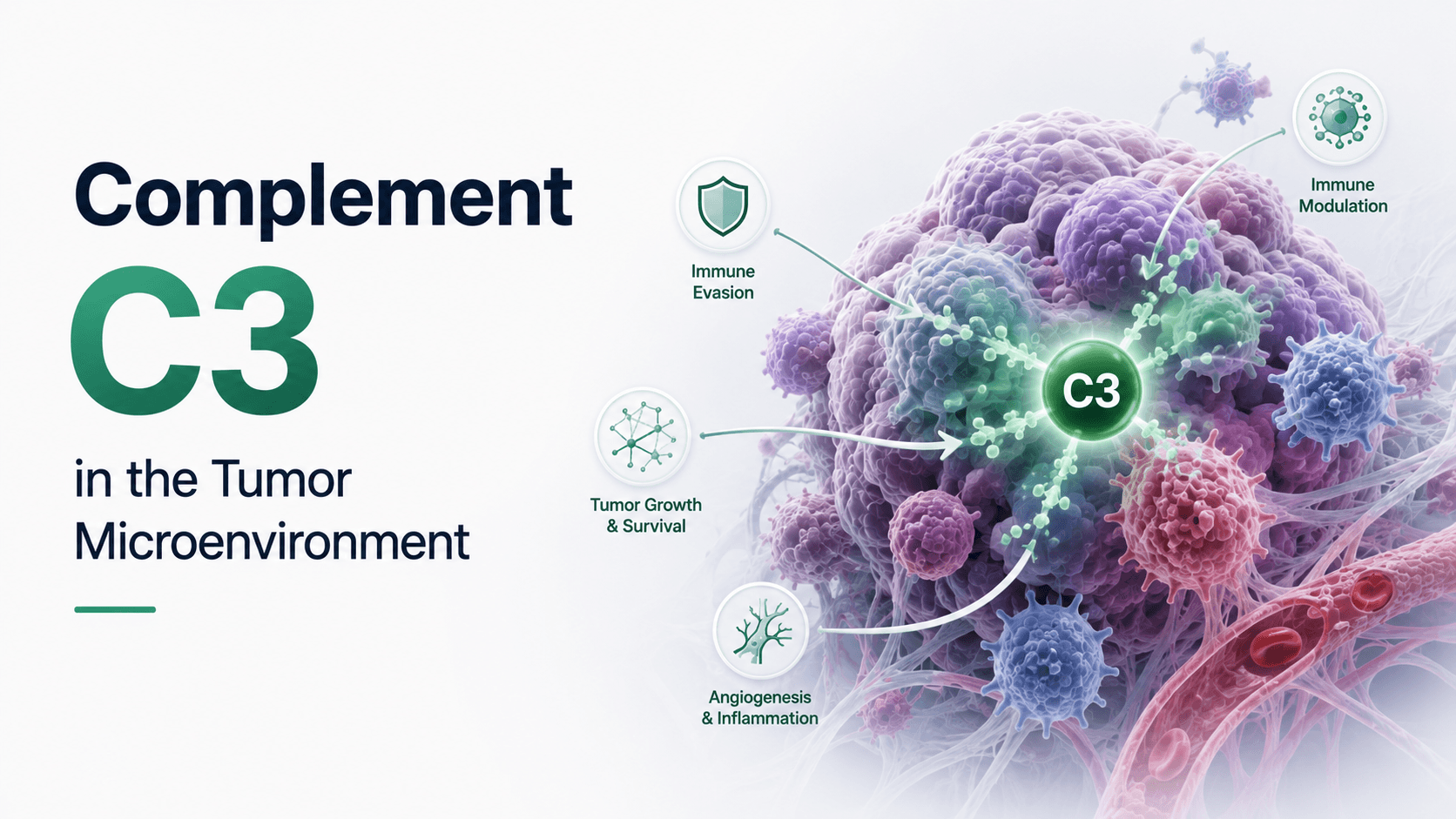
For years, complement C3 has been filed away as a liver-synthesized acute-phase protein whose main job is opsonizing pathogens in the bloodstream - relevant to sepsis and autoimmunity, not oncology. That framing is now incomplete. Researchers at Nagoya University, publishing in Nature Communications, found that C3 produced locally by cancer-associated fibroblasts inside the tumor microenvironment determines whether immune checkpoint blockade works, while circulating C3 levels made essentially no difference to outcomes. This distinction - tumor-produced versus liver-produced C3 - matters directly to hepatology, because the liver is both the primary site of systemic C3 synthesis and, in HCC, the organ where this local immune mechanism plays out. For a subspecialty already managing the first-line complement C3 hepatocellular carcinoma immunotherapy question through atezolizumab-bevacizumab, a locally-acting biomarker that blood tests won't detect changes what "checking the immune status of a tumor" should mean.
How Local Complement C3 Shapes the Tumor Microenvironment
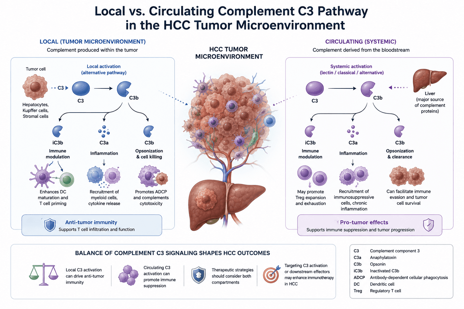
The mechanism runs through myeloid cell trafficking, not T-cell priming. When C3 produced by fibroblasts within the tumor breaks down, it generates a fragment called iC3b, which blocks immunosuppressive myeloid cells from infiltrating the tumor stroma. Fewer suppressive myeloid cells in the tumor microenvironment HCC means checkpoint inhibitors have a clearer path to reactivating exhausted T cells.
The research team confirmed the source-specificity experimentally in mice: reducing liver-derived circulating C3 by 90% barely touched anti-PD-1 efficacy, while silencing fibroblast-derived C3 within the tumor - despite only a 9% drop in blood C3 - sharply reduced how well the same antibody worked. That asymmetry is the finding worth sitting with. A hepatologist reflexively thinks of C3 as a liver synthesis product, and in the systemic sense it is. But the protein doing the immunologically relevant work in the tumor is made on-site, by fibroblasts, and behaves independently of whatever the liver is putting into circulation. Per the study, tumor tissue from lung cancer patients showed the same pattern clinically: roughly half of patients with high intratumoral C3 responded to immunotherapy, versus none of those with low levels - while blood C3 tracked with nothing.
Case in Point
A 58-year-old woman with MASH-related HCC, Child-Pugh A, BCLC stage B, is started on atezolizumab-bevacizumab after declining for locoregional therapy alone. Her baseline complement panel, drawn as part of a broader metabolic workup, shows normal serum C3. Nine months later, imaging shows progression despite an unremarkable inflammatory profile and no obvious resistance mechanism on standard workup.
Retrospectively, this is exactly the scenario the Nagoya group's data would predict poorly: a normal systemic C3 tells you nothing about what her cancer-associated fibroblasts were producing locally. If tumor-tissue C3 or iC3b immunohistochemistry becomes clinically available, a case like hers is where it would earn its place - not as confirmation after the fact, but as a pre-treatment stratifier that a serum complement panel could never provide, since the two compartments dissociate almost entirely.
What This Could Mean for HCC Treatment Selection
None of this changes today's first-line regimen - atezolizumab-bevacizumab remains standard of care per IMbrave150, and no tissue-based complement assay has cleared validation for clinical use. What it does is open a biomarker pathway distinct from PD-L1 expression, tumor mutational burden, or the immune-based HCC subtyping schemes already in circulation, none of which have reliably predicted response at the individual-patient level.
The mouse data go further than description: the research team tested a drug that mimics iC3b's myeloid-blocking effect and found it restored immunotherapy efficacy in tumors that had previously resisted treatment, extending survival significantly. That is the more consequential implication for immune checkpoint inhibitor resistance in HCC - not a better test to identify non-responders, but a potential add-on strategy to convert them into responders. For a disease where second-line options after checkpoint failure remain thin, a mechanism-matched combination agent, if it clears human trials, would be the more practice-changing outcome of the two. A tissue-based immunotherapy biomarker liver cancer assay is the more immediate translational step; the myeloid-blocking drug is the one worth watching over the next several years.
A Frequently Overlooked Point
The instinct in hepatology is to treat complement as a systemic marker of liver synthetic function - worth checking in the context of cirrhosis, sepsis, or autoimmune workups, and otherwise not particularly actionable in oncology. This data set argues that instinct now has a blind spot. iC3b myeloid cells trafficking is a tumor-local event, produced by stromal cells that have nothing to do with hepatic synthetic reserve, and a normal or even elevated serum complement panel provides no visibility into it. Until tissue-based assays exist, the honest clinical position is that we currently have no reliable way to know a given patient's intratumoral complement status - which is itself useful to communicate to a patient asking why the same drug worked for someone else and not for them.
Bottom Line for Clinical Practice
Serum complement C3 does not reflect intratumoral C3 activity - do not use a systemic complement panel to infer immunotherapy responsiveness in HCC.
Tumor-produced C3, broken down locally into iC3b, blocks immunosuppressive myeloid cell infiltration and appears to be what determines checkpoint inhibitor efficacy at the tissue level.
No validated clinical assay for intratumoral C3 or iC3b currently exists; atezolizumab-bevacizumab remains first-line per IMbrave150 regardless of complement status.
Watch for translational work on iC3b-mimetic agents as a potential combination strategy for atezolizumab bevacizumab response in patients who progress on checkpoint blockade alone.
When a patient's HCC isn't responding the way the literature says it should, the next question is rarely simple - and increasingly, the answer sits in the tumor microenvironment, not the chart. Walk GastroAGI through a case like this one, and it'll help you reason through what's actually driving non-response, guideline-anchored and current.
Related Blog: GVAX Pancreatic Cancer Vaccine: What the 2026 Trial Data Means for PDAC Care
Also Read: APASL 2026 Istanbul: Key Clinical Takeaways Every Hepatologist Needs to Know


We are pioneers in clinical intelligence, dedicated to helping gastroenterologists harness the power of artificial intelligence to drive precision, efficiency, and patient growth.