
Can EGGIM Reliably Identify Extensive Gastric Intestinal Metaplasia? New Meta-analysis Supports Real-Time Endoscopic Risk Stratification
GIE meta-analysis found EGGIM ≥5 highly accurate for detecting extensive gastric intestinal metaplasia using NBI or BLI endoscopy.


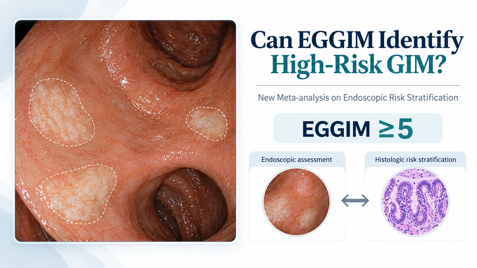
Can Endoscopy Identify High-Risk Gastric Intestinal Metaplasia Before Histology Does?
For clinicians evaluating gastric intestinal metaplasia, the central challenge is not simply identifying metaplasia. It is determining its extent well enough to identify patients who may warrant closer surveillance.
Histologic staging using the Operative Link on Gastric Intestinal Metaplasia, or OLGIM, provides one framework for this risk stratification. However, OLGIM depends on biopsy sampling and subsequent histopathologic assessment. The possibility of estimating the extent of gastric intestinal metaplasia during the endoscopic examination itself is therefore clinically attractive.
The Endoscopic Grading of Gastric Intestinal Metaplasia, or EGGIM, was developed as a real-time endoscopic staging approach. Rather than waiting for histology to define the extent of metaplasia, EGGIM uses endoscopic assessment of different gastric regions to generate a score reflecting the distribution of intestinal metaplasia.
A new systematic review and meta-analysis by Luu and colleagues, published online in Gastrointestinal Endoscopy on September 1, 2026, examined how accurately EGGIM identifies extensive gastric intestinal metaplasia when histologic OLGIM staging is used as the reference standard. The analysis specifically focused on advanced imaging approaches using narrow-band imaging or blue light imaging.
The findings are encouraging: an EGGIM score of at least 5 showed high pooled sensitivity and specificity for identifying OLGIM stage III–IV disease. But the study should be interpreted as evidence supporting diagnostic accuracy, not as proof that EGGIM can replace histology or independently determine surveillance strategy.
The Clinical Problem Is Extent, Not Merely Presence
Gastric intestinal metaplasia is a precancerous gastric condition, but its clinical significance is not uniform across all patients. One of the key distinctions is whether metaplastic change is limited or extensive.
The meta-analysis defined extensive gastric intestinal metaplasia as OLGIM stage III–IV. This is an important design feature because the investigators were not asking whether EGGIM can detect any focus of intestinal metaplasia. Instead, they evaluated whether an endoscopic score could discriminate patients with more extensive histologic involvement from those without it.
That question has practical implications. Histologic risk stratification requires biopsies to be taken from relevant gastric sites, processed, and interpreted. Endoscopic grading has the potential to provide immediate information during the examination and may help the endoscopist conceptualize the distribution of gastric intestinal metaplasia before pathology results are available.
However, diagnostic usefulness depends on accuracy. A staging system that frequently misses extensive disease would be unsafe as a risk-stratification tool, while one with poor specificity could classify too many patients as having advanced disease. Sensitivity and specificity are therefore central to judging whether EGGIM has practical potential.
How the Meta-analysis Was Designed
Luu and colleagues performed a systematic review and diagnostic accuracy meta-analysis. They searched PubMed, Embase, Scopus, the Cochrane Library, and ClinicalTrials.gov through February 2026. Eligible studies enrolled adults undergoing upper gastrointestinal endoscopy and assessed EGGIM against histologic OLGIM staging as the reference standard.
The authors identified nine studies overall. For the primary pooled analysis, they restricted inclusion to seven studies involving 1,143 patients in which EGGIM was performed using narrow-band imaging or blue light imaging. All seven studies used an EGGIM cutoff of at least 5 to identify extensive gastric intestinal metaplasia.
This restriction is clinically relevant. Modern image-enhanced endoscopy can improve visualization of mucosal and vascular patterns compared with conventional white-light imaging alone. By limiting the primary analysis to NBI- or BLI-compatible approaches, the investigators examined EGGIM within the technological context in which it is most likely to be used as a structured endoscopic staging tool.
The statistical analysis used a hierarchical bivariate random-effects model to pool sensitivity and specificity. The investigators also calculated likelihood ratios, diagnostic odds ratio, and the area under the summary receiver operating characteristic curve. Subgroup analyses explored differences according to imaging modality and geographic region.
What EGGIM ≥5 Actually Achieved
The principal result was a pooled sensitivity of 0.90, with a 95% confidence interval of 0.84 to 0.95, for identifying extensive gastric intestinal metaplasia. Pooled specificity was 0.92, with a 95% confidence interval of 0.89 to 0.94.
In practical diagnostic terms, the pooled data suggest that an EGGIM threshold of at least 5 correctly identified a high proportion of patients with OLGIM stage III–IV disease while also correctly classifying most patients without extensive disease.
The diagnostic odds ratio was 105.93, with a 95% confidence interval of 53.81 to 208.54. The area under the summary receiver operating characteristic curve was 0.96, with a 95% confidence interval of 0.94 to 0.97.
An AUC close to 1 indicates strong overall discrimination between patients with and without the target condition. In this context, the reported AUC supports the ability of EGGIM ≥5, when used with NBI or BLI, to distinguish extensive histologic gastric intestinal metaplasia from less extensive disease.
These numbers are strong for a diagnostic staging tool. They are also consistent with the authors’ conclusion that EGGIM appears to have high diagnostic accuracy for identifying extensive gastric intestinal metaplasia.
Yet diagnostic accuracy is only one part of clinical utility. A test can perform well against a reference standard and still require further evidence before it changes how patients are managed.
Why Image-Enhanced Endoscopy Matters Here
The primary analysis was deliberately limited to studies using NBI or BLI. That makes the study more specific than a general assessment of visual recognition of intestinal metaplasia.
EGGIM is intended to stage metaplasia according to its endoscopically visible extent. Reliable recognition therefore depends on being able to distinguish mucosal patterns associated with gastric intestinal metaplasia across multiple gastric regions.
The current meta-analysis supports EGGIM specifically in the setting of image-enhanced endoscopy compatible with narrow-band imaging or blue light imaging. Clinicians should therefore avoid extrapolating the pooled performance directly to endoscopic examinations performed without these modalities.
The authors also examined performance according to NBI versus BLI. In descriptive subgroup analyses, diagnostic performance appeared broadly similar between the two platforms.
This is potentially important because it suggests that the EGGIM concept may not depend on a single image-enhancement platform. However, the authors did not perform a formal statistical comparison between these subgroups because the number of studies was limited. The apparent similarity should therefore be considered descriptive rather than proof of equivalence.
Eastern and Western Studies: Similar Performance Despite Different Prevalence
Another notable component of the meta-analysis was the comparison between Eastern and Western study populations.
The prevalence of extensive gastric intestinal metaplasia differed substantially between the geographic groups. It was 55% in Eastern studies compared with 18% in Western studies, a statistically significant difference with P < .001.
Despite this difference in disease prevalence, the diagnostic performance of EGGIM appeared broadly similar between Eastern and Western studies in descriptive subgroup analysis.
This finding is potentially reassuring for generalizability. A diagnostic system that performs consistently across populations with different baseline prevalence would be attractive for broader use.
However, caution is needed. The authors explicitly note that formal between-subgroup comparisons were not performed because the number of available studies was limited. Therefore, the meta-analysis does not establish that EGGIM performs identically across all geographic settings.
Prevalence also has practical implications beyond sensitivity and specificity. Even when these test characteristics remain relatively stable, the probability that a positive or negative result reflects true disease can vary according to underlying disease prevalence. Clinicians should therefore interpret any endoscopic staging system within the clinical context of the population being examined.
A Potential Shift Toward Real-Time Gastric Risk Stratification
The most clinically interesting aspect of EGGIM is immediacy.
Histologic staging requires tissue acquisition and pathology. EGGIM offers an endoscopic estimate during the procedure itself. If the distribution of gastric intestinal metaplasia can be accurately characterized in real time, the endoscopist gains additional information while still examining the stomach.
This may eventually influence how endoscopists think about mapping, targeted inspection, documentation, or subsequent surveillance planning. However, those downstream applications were not tested by this meta-analysis.
The study evaluated diagnostic accuracy against OLGIM stage III–IV. It did not test whether EGGIM-guided management improves gastric cancer detection, reduces mortality, decreases unnecessary biopsies, lowers cost, or safely replaces standardized histologic staging.
The appropriate conclusion is therefore narrower: EGGIM ≥5 appears to identify extensive GIM with high diagnostic accuracy when used with NBI or BLI.
That is an important finding, but it remains a diagnostic finding.
What the Study Does Not Establish
The strength of the pooled sensitivity and specificity may tempt clinicians to ask whether EGGIM could replace biopsy-based OLGIM staging. The current evidence does not answer that question.
OLGIM was the reference standard in the included studies. EGGIM was evaluated by how closely it identified disease defined histologically. Therefore, the analysis validates EGGIM against pathology rather than demonstrating that pathology is unnecessary.
Similarly, the study does not establish that an EGGIM score of at least 5 should independently trigger surveillance or determine surveillance intervals. Surveillance recommendations involve more than diagnostic accuracy alone and may incorporate patient-level risk factors, histology, family history, Helicobacter pylori status, and guideline-specific considerations. Those questions were outside the scope of this meta-analysis.
The study also does not demonstrate that EGGIM reduces gastric cancer incidence or improves survival.
Clinicians should distinguish three separate propositions:
EGGIM may accurately identify extensive histologic GIM.
EGGIM may ultimately become useful in risk stratification.
EGGIM alone can direct surveillance or replace histologic assessment.
The meta-analysis provides substantial support for the first proposition and encouraging evidence for the second. It does not establish the third.
Strengths of the Evidence
Several features strengthen the study.
First, it used a clearly defined reference standard: histologic OLGIM stage III–IV. Second, the authors focused the primary pooled analysis on modern image-enhanced endoscopy using NBI or BLI. Third, all seven studies in the primary analysis used the same EGGIM threshold of at least 5, reducing one important source of threshold heterogeneity.
The study also used statistical methods appropriate for pooling diagnostic accuracy data and reported multiple complementary performance measures rather than sensitivity or specificity alone.
Finally, the analysis explored potential differences by geography and imaging platform, both of which are highly relevant to real-world implementation.
Where Uncertainty Remains
The authors themselves emphasize that additional studies are needed to confirm the findings and strengthen the evidence base.
One limitation is simply the number of available studies. Although 1,143 patients were included in the primary pooled analysis, only seven NBI/BLI studies contributed to that analysis. This limited the ability to perform robust formal comparisons across important subgroups.
The geographic and imaging-platform analyses were therefore descriptive. Similar apparent performance between East and West or between NBI and BLI should not be interpreted as definitive equivalence.
Another issue is implementation. A diagnostic grading system is only as reliable as the endoscopist applying it. The published abstract does not establish how much training is required, whether accuracy differs by operator experience, or how reproducible EGGIM is when used outside study environments.
Those are crucial questions if EGGIM is to move from research validation into widespread routine practice.
Where Future Research Should Go
The next phase of research should move beyond confirming sensitivity and specificity.
Prospective studies can examine how EGGIM performs when applied across diverse endoscopy units, operators, patient populations, and imaging platforms. Reproducibility between endoscopists will be important, particularly if the score is intended for routine risk stratification.
Studies should also determine whether incorporating EGGIM into clinical pathways changes management in a beneficial way.
For example, can EGGIM improve the efficiency or targeting of biopsy protocols without compromising diagnostic certainty? Can it help identify patients who warrant closer surveillance? Does it add meaningful information beyond histology and established clinical risk factors? And most importantly, does an EGGIM-guided strategy ultimately improve clinically relevant outcomes?
Those questions remain unanswered by the current meta-analysis.

Clinical Takeaway
The 2026 Gastrointestinal Endoscopy systematic review and meta-analysis provides strong diagnostic evidence supporting EGGIM as a real-time endoscopic tool for identifying extensive gastric intestinal metaplasia.
Across seven NBI/BLI studies involving 1,143 patients, an EGGIM score of at least 5 showed pooled sensitivity of 90% and specificity of 92% for detecting OLGIM stage III–IV disease. The summary AUC was 0.96, indicating high overall discriminatory performance.
The results support the validity of EGGIM as an endoscopic risk-stratification approach, particularly when image-enhanced endoscopy is used. They do not, however, establish that EGGIM should replace histologic staging, determine surveillance intervals on its own, or be considered an independent surrogate for future gastric cancer outcomes.
For gastroenterologists and endoscopists, the practice signal is therefore promising but measured: EGGIM ≥5 appears to be a highly accurate marker of extensive gastric intestinal metaplasia, but its optimal role within surveillance and biopsy strategies still requires prospective validation.
Five key clinical takeaways
This was a systematic review and diagnostic accuracy meta-analysis, published online in Gastrointestinal Endoscopy on September 1, 2026. The investigators evaluated EGGIM against histologic OLGIM staging.
Seven NBI/BLI studies involving 1,143 patients contributed to the primary pooled analysis, and all used an EGGIM cutoff of ≥5 to identify extensive GIM.
EGGIM ≥5 showed high diagnostic accuracy for OLGIM stage III–IV disease, with pooled sensitivity of 0.90 and specificity of 0.92.
Overall discrimination was strong, with a diagnostic odds ratio of 105.93 and summary ROC AUC of 0.96. Performance appeared broadly similar across NBI and BLI and Eastern and Western studies, although formal subgroup comparisons were not performed.
The study supports EGGIM as a real-time endoscopic risk-stratification tool but does not establish it as a replacement for histology or as an independent surveillance algorithm. Further validation and implementation studies remain necessary.
Source
Luu MN, Trinh NA, Tran TLT, Dang TP, Hiyama T, Quach DT. Diagnostic Accuracy of the Endoscopic Grading of Gastric Intestinal Metaplasia for Detecting Extensive Disease: a Systematic Review and Meta-analysis. Gastrointestinal Endoscopy. Published online September 1, 2026. DOI: 10.1016/j.gie.2026.08.036. PMID: 42679864.
References

We are pioneers in clinical intelligence, dedicated to helping gastroenterologists harness the power of artificial intelligence to drive precision, efficiency, and patient growth.