GastroAGI Logo
OverviewBlogsAbout
Trending TopicsDaily BriefConference

Gastroenterology Insights — Clinical Updates, Guides & Resources — Page 11

Top Gastroenterology Conferences in India: ISGCON, INASL, SGEI & WCOG 2026
7 min readApr 15, 20260reads

Top Gastroenterology Conferences in India: ISGCON, INASL, SGEI & WCOG 2026

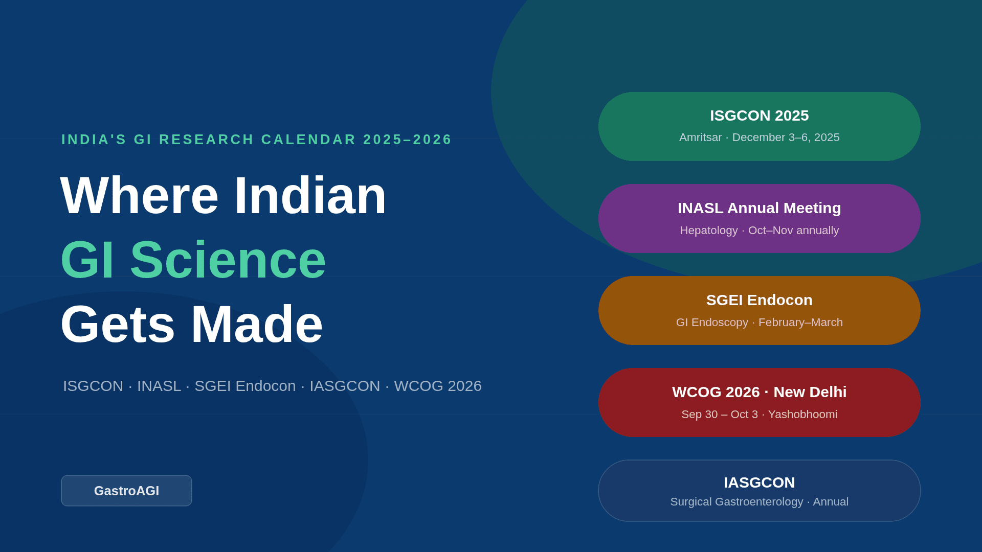
A DM physician at a tier-2 hospital spends hours each week parsing conflicting treatment protocols for NAFLD — one from a US guideline body, another from a European liver society. Neither accounts for Indian dietary patterns, genetic predispositions, or the public-health context of a country carrying nearly 20% of the world's chronic liver disease burden. The answer, most of the time, isn't in a journal — it's on a conference floor in Varanasi, Amritsar, or New Delhi, where Indian gastroenterologists are producing and debating exactly that evidence. This post maps every major national conference where that work happens. India's gastroenterology disease burden is distinctive. Helicobacter pylori prevalence remains above 50% in many populations. Non-alcoholic fatty liver disease is accelerating alongside the metabolic syndrome epidemic. Inflammatory bowel disease is rising sharply in urban cohorts, yet presentation patterns diverge from Western populations in ways that make direct guideline application unreliable. Research on these nuances does not emerge from single-centre case reports — it emerges from the multi-institutional collaborations, abstract presentations, and consensus workshops that national conferences make possible. For Indian gastroenterologists, residents, and researchers, knowing which conferences matter — and what each one offers — is not a scheduling convenience. It is a core part of staying current and contributing to evidence that is clinically relevant to Indian patients. The landscape of gastroenterology conferences in India is richer than many outside the field realise, spanning medical gastroenterology, hepatology, surgical gastroenterology, and advanced endoscopy as distinct but interlinked domains. The National Pillars: ISGCON, INASL, and SGEI Endocon Three societies form the backbone of organised gastroenterology in India, and their annual or semi-annual meetings function as the primary venues for disseminating Indian GI research. ISGCON — Annual Congress of the Indian Society of Gastroenterology Organised by: Indian Society of Gastroenterology (ISG) · Over 4,000 members across 21 state chapters Annual: December ISGCON is the flagship national event for medical gastroenterology in India. Held every December, the 65th edition convened in Varanasi in December 2024, and the 66th — ISGCON 2025 — was organised in Amritsar from 3–6 December 2025, with 2,000–2,500 delegates and both national and international faculty. The conference publishes its abstracts in a dedicated supplement of the Indian Journal of Gastroenterology , making it a PubMed-indexed source of Indian GI research. Symposia, live procedure demonstrations, orations, and best-paper competitions give the meeting both educational and competitive depth. The P.N. Chhuttani Distinguished Achievement Oration and the R.K. Tandon Oration are among the most prestigious recognitions in Indian academic gastroenterology — both awarded at ISGCON. INASL Annual Meeting — Indian National Association for Study of the Liver Organised by: INASL · Focused on hepatology and liver disease Annual: Mid-term meetings INASL runs a full annual scientific meeting alongside several mid-term and subspecialty events each year. The joint national meeting with ISG and SGEI — typically held in October-November — is the largest single gathering for hepatologists and gastroenterologists in India in the autumn calendar. In 2024, the 32nd Annual Scientific Meeting was held in Kochi. INASL's upcoming calendar includes the Bengal Liver Summit (January 2026, Kolkata), Grand Rounds in Hepatology (May 2026, Mumbai), and the INASL-ILCA School of Liver Cancer. Each meeting generates symposia and consensus statements directly relevant to Indian liver disease management. SGEI Endocon — Society of Gastrointestinal Endoscopy of India Organised by: SGEI · ~1,950 members · Advanced endoscopy focus Two meetings/year : Live workshops SGEI holds two annual meetings. The first, typically in February-March, is a two-day live endoscopy workshop covering both basic and advanced procedures — EUS, ERCP, ESD, POEM, and endo-bariatric techniques. The second is the joint national meeting with ISG and INASL. SGEI collaborates with the World Endoscopy Organisation (WEO), OMED, and the American Society of GI Endoscopy (ASGE), and regularly hosts international endoscopists for interactive sessions. For trainees, the SGEI workshop circuit is among the most practical skill-building opportunities available in the country. Case in point A second-year DM Gastroenterology resident presents an abstract at ISGCON on the prevalence of H. pylori clarithromycin resistance in a tertiary centre cohort from eastern India — data that directly challenges the standard first-line triple therapy recommendations still cited in most institutional protocols. The abstract is accepted for an oral presentation in the clinical research session. The presentation prompts a floor discussion with investigators from three other centres who have documented similar resistance trends. By the end of the conference, the resident has been invited to join a multicentre collaborative audit — the kind of collaboration that turns a single-centre observation into nationally generalisable evidence. This is the specific mechanism by which ISGCON generates research that reshapes Indian clinical practice, and why conference participation for residents is not extracurricular — it is formative. Beyond the Big Three: IASGCON, State Chapters, and WCOG 2026 IASGCON — Indian Association of Surgical Gastroenterology Organised by: IASG (a section of Association of Surgeons of India) · Founded 1988 Annual : Surgical GI focus IASG focuses exclusively on surgical gastroenterology — laparoscopic procedures, hepatopancreaticobiliary surgery, GI oncology, and bariatric surgery. IASGCON draws surgeons with subspecialty interest in the GI tract, and the annual meeting features live surgical demonstrations, debates on management controversies (e.g., timing of cholecystectomy in acute cholecystitis, surgical vs. endoscopic management of common bile duct stones), and postgraduate clinical courses. For gastroenterologists managing complex patients who will need surgical input, understanding the surgical gastroenterology literature that emerges from IASGCON is directly relevant to shared decision-making. WCOG 2026 — World Congress of Gastroenterology · New Delhi Venue: Yashobhoomi Convention Centre, New Delhi · Dates: 30 September – 3 October 2026 Global : Landmark The World Congress of Gastroenterology returns to India in 2026 — a landmark event that will position Indian GI research on the global stage. Hosted at Yashobhoomi in New Delhi from 30 September to 3 October 2026, WCOG 2026 will draw gastroenterologists, hepatologists, endoscopists, and researchers from across the world. For Indian clinicians and researchers, this is a once-in-a-generation opportunity to present work to an international audience, engage with cutting-edge global protocols, and establish collaborations that influence Indian research priorities for years ahead. State chapter meetings — including TNISGCON (Tamil Nadu), the Bengal Liver Summit, and ISGKCON (Karnataka) — are where regional disease patterns and local practice variations receive dedicated attention. These smaller, focused meetings often generate the hypothesis-forming data that subsequently appears in national conference abstracts and peer-reviewed publications. A frequently overlooked point about conference engagement Most gastroenterologists attend conferences for the plenary sessions and live demonstrations — the visible content. The less visible but arguably more valuable output is the informal consensus that forms around contested clinical questions: How long should bismuth quadruple therapy run in a high-resistance setting? At what platelet count do experienced Indian hepatologists actually scope a patient with cirrhosis? What are the real complication rates of ESD at Indian volume centres? These conversations happen in hallways, at dinner tables, and in breakout workshops — they don't appear in the abstract supplements. This is why the argument for attending rather than just reading the proceedings is not about prestige. It's about accessing a layer of clinical reasoning that never makes it to print. Bottom line for clinical practice ISGCON every December is the single most important date on the Indian gastroenterology calendar — abstract submission opens months in advance and the proceedings are PubMed-indexed via the Indian Journal of Gastroenterology . INASL's mid-term meetings (Bengal Liver Summit, Grand Rounds in Hepatology) are not smaller versions of the annual meeting — they are specialty-dense, focused events often better for hepatologists than the larger joint congress. SGEI's February-March live workshop is the highest-yield skills event for trainees in therapeutic endoscopy — prioritise it before seeking international hands-on courses. WCOG 2026 in New Delhi is a once-in-a-generation home-ground opportunity to establish international research collaborations. Begin preparing abstracts now. State chapter meetings (TNISGCON, Bengal Liver Summit, ISGKCON) are where the hypothesis-generating regional data lives — they deserve a place in the annual calendar alongside the national meetings. Before you walk into your next conference, let GastroAGI help you map the clinical questions worth pursuing — from IBD phenotyping to H. pylori resistance patterns. Walk the AI through your patient caseload, and it will return guideline-anchored reasoning and research angles in seconds. Go in with a question worth answering.

GastroAGI TeamRead more
Fatigue in Aging: When to Stop Reassuring and Start Investigating
8 min readApr 14, 202640reads

Fatigue in Aging: When to Stop Reassuring and Start Investigating

Fatigue in older adults is one of the most dismissed symptoms in medicine — and one of the most dangerous to ignore. Nearly one-third of people over 65 report significant fatigue , yet it's routinely chalked up to "just getting older." That assumption costs people years of quality life. Persistent fatigue is a signal, not a sentence. The biological mechanisms driving exhaustion in aging are increasingly well-understood — and many are treatable. Understanding where to draw the line between normal tiredness and a symptom worth pursuing starts at the cellular level. Mitochondrial Dysfunction and Chronic Inflammation: The Two Strongest Biological Links Age-related fatigue isn't simply about doing less or sleeping poorly — it has measurable biological roots. Two mechanisms dominate the research: declining mitochondrial efficiency and low-grade chronic inflammation, sometimes called "inflammaging." According to a multi-dimensional fatigue model published in PMC , these converging processes erode energy production at the cellular level, making fatigue in older adults physiologically distinct from ordinary tiredness. Fatigue driven by biology demands a biological investigation — not reassurance. Understanding this distinction is exactly what shapes the clinical picture explored next. Clinical Scenario Understanding the full range of elderly fatigue causes requires more than a list of diagnoses — it helps to see how they converge in one person. Consider a 74-year-old woman whose physician keeps attributing her exhaustion to "getting older." Meanwhile, unmanaged hypothyroidism, mild anemia, and disrupted sleep quietly compound each other. As research from the NIA confirms, fatigue in older adults frequently has multiple, treatable contributors hiding behind a single vague complaint. Recognizing that overlap is exactly where rigorous clinical investigation begins. When Fatigue Is a Geroscience Signal: The Clinical Investigation Framework Tiredness in aging rarely arrives with a single, clean explanation. What the clinical scenario above illustrates is that fatigue often functions as a convergence signal — one symptom reflecting several interacting systems simultaneously losing ground. Research from Frontiers in Psychology confirms that fatigue patterns shift meaningfully across aging trajectories, making longitudinal awareness essential. Persistent, unexplained exhaustion deserves structured investigation — not dismissal. That investigation framework typically examines three domains: metabolic function, inflammatory burden, and physiological reserve. When all three are compromised, fatigue compounds rapidly — something routine reassurance simply can't address. A Frequently Overlooked Point: The Reassurance Trap Chronic fatigue in older adults is routinely dismissed as an inevitable part of aging — and that dismissal has real consequences. A common pattern is for clinicians and caregivers alike to offer reassurance without investigation, effectively normalizing what may be an actionable signal. Research from Frontiers in Psychology confirms fatigue trajectories in aging are far from uniform — meaning "it's just age" is rarely the complete answer. Unchallenged reassurance doesn't comfort patients; it delays care. Bottom Line for Clinical Practice Fatigue in seniors is not a footnote — it's a signal. As the previous sections have made clear, defaulting to reassurance without investigation carries real clinical risk. In practice, persistent fatigue warrants a structured response: rule out treatable causes first, acknowledge complexity second, and never let "it's just aging" close the conversation prematurely. The next section explores what that investigation actually looks like in older adults day to day. Fatigue in Older Adults Fatigue doesn't affect all seniors equally. Research shows that fatigue in elderly women is reported at notably higher rates than in men of the same age — a pattern linked to hormonal changes, higher rates of autoimmune conditions, and differences in how women report symptoms. According to Cleveland Clinic , this disparity makes gender an important variable when evaluating unexplained tiredness. Recognizing these demographic nuances is exactly the kind of detail that separates thorough clinical thinking from blanket reassurance — and it connects directly to the guidance that major health institutions have formalized on this topic. National Institute on Aging Energy levels aging is a topic the National Institute on Aging takes seriously — and their guidance reinforces what clinicians are seeing in practice. The NIA's dedicated resource on fatigue in older adults emphasizes that persistent tiredness warrants evaluation, not dismissal. Fatigue that limits daily function is a clinical concern , full stop — and federal health authorities agree. Why Am I Tired? What NIH Research Tells Us About Fatigue in Older Adults Asking "why am I tired" is one of the most common — and most underexplored — questions in senior health. Hebrew SeniorLife's overview of fatigue in older adults reinforces a key clinical reality: tiredness in seniors is rarely one-dimensional. It intersects physical health, mental well-being, and social engagement simultaneously. Fatigue that persists deserves investigation, not reassurance. That distinction matters — and it sets the stage for understanding exactly when age-related tiredness crosses a line worth examining. At What Age Do You Start Feeling Really Tired? Fatigue doesn't arrive on a fixed birthday. Research published in Frontiers in Psychology shows fatigue increases gradually across adulthood, accelerating noticeably after 60. However, hypothyroidism fatigue can mimic or amplify age-related tiredness at virtually any age — making it a condition worth ruling out before attributing exhaustion purely to getting older. What Is Chronic Fatigue Syndrome? Chronic fatigue syndrome (CFS) — also called myalgic encephalomyelitis (ME/CFS) — is a distinct, diagnosable condition, not simply feeling run-down. It's characterized by extreme exhaustion that doesn't improve with rest, lasts six months or longer, and significantly limits daily activity. Unlike typical age-related tiredness, CFS often worsens with physical or mental exertion — a hallmark symptom known as post-exertional malaise . Recognizing this distinction matters, because CFS requires targeted investigation rather than reassurance. What Helps Fatigue? Addressing fatigue effectively starts with identifying its root cause — which is exactly why earlier sections emphasized investigation over reassurance. Once underlying conditions are treated, several evidence-based strategies consistently make a real difference: Regular, moderate exercise — counterintuitively, gentle movement reduces fatigue over time Sleep hygiene improvements — consistent sleep schedules and limiting daytime napping Nutritional support — correcting deficiencies in iron, B12, or vitamin D Medication review — eliminating or adjusting drugs that cause drowsiness Understanding why these strategies work at a biological level, though, is a question worth exploring next. ELI5: Why Do You Become More Tired the Older You Become? Think of your body like a car engine with slowly accumulating wear. Muscle mass decreases, mitochondria (your cells' energy generators) become less efficient, and the cardiovascular system works harder to deliver oxygen. Energy production quietly declines while energy demands don't. The result? Everyday tasks cost more effort than they once did — and that accumulating gap is what aging fatigue actually feels like. That biological reality sets the stage for an interesting question: why do some older adults still radiate vitality while others seem perpetually drained? How Do Some Older People Still Look "Awake" While Others Don't? Lifestyle factors explain much of this gap. Older adults who maintain regular physical activity, strong social connections, consistent sleep schedules, and well-managed chronic conditions tend to preserve significantly more energy. Cumulative habits — not genetics alone — drive much of the visible difference. That vitality you notice in some people isn't luck; it's usually decades of sustainable choices quietly adding up. This sets the stage for a deeper question: at what point does increasing tiredness cross from normal aging into something worth investigating? Is It Normal to Start Feeling Tired More Often Once You Reach a Certain Age? Yes — to a point. Some increase in fatigue is a predictable part of aging, driven by the biological and lifestyle shifts covered earlier in this article. Feeling less energetic than you did at 35 is expected at 65. However, "normal" has real limits, and fatigue that disrupts daily functioning deserves attention rather than automatic reassurance. That distinction matters more than most people realize. Why Do Older People Often Feel More Tired, and Could There Be More to It? Fatigue in older adults rarely has a single cause. Biology, lifestyle, and underlying health conditions typically combine — making it easy to dismiss what deserves a closer look. Cumulative fatigue often signals something worth investigating, not just accepting. As the next section explores, that distinction becomes especially important when fatigue starts affecting daily function. Is It Common for Older Individuals to Experience Fatigue? Yes — fatigue is remarkably prevalent among older adults. Research consistently shows that fatigue affects between 40% and 74% of older adults , making it one of the most frequently reported complaints in this population. It's common, but "common" doesn't mean it should be dismissed or accepted without question. That distinction matters enormously when deciding whether to act — a point worth keeping in mind as you consider what persistent tiredness at any age might actually be signaling. I Am 75 Years Old and Feel Tired a Lot. What Should I Do? Persistent tiredness at 75 isn't something to simply accept. Start by tracking your fatigue — note when it peaks, what worsens it, and whether rest helps. Then bring those observations to your doctor. As the National Institute on Aging advises, unexplained fatigue always warrants a medical evaluation to rule out treatable causes like anemia, thyroid issues, or medication side effects. This applies especially to women, whose experience of fatigue often carries distinct patterns worth examining closely. Fatigue in Elderly Women Women over 65 often report fatigue at higher rates than their male peers, with hormonal shifts, higher rates of autoimmune conditions, and greater likelihood of depression all contributing. Fatigue in elderly women deserves gender-specific attention — what looks like general tiredness may reflect thyroid dysfunction, anemia, or lingering effects of menopause-related sleep disruption. These patterns warrant targeted investigation rather than blanket reassurance. For some older women, persistent, unexplained exhaustion points toward something more complex — which brings us to an important distinction explored next. What Is Chronic Fatigue Syndrome? Persistent, unexplained exhaustion that doesn't improve with rest deserves serious medical attention — not reassurance. Throughout this article, one truth stands out: fatigue in aging is never just "getting old." From hormonal shifts to sleep disorders, the causes are real, treatable, and worth investigating. Talk to your doctor. Don't wait.

GastroAGI TeamRead more
Why the 6-Month Rule No Longer Holds Up | Early Liver Transplantation for Alcoholic Hepatitis:
6 min readApr 9, 202653reads

Why the 6-Month Rule No Longer Holds Up | Early Liver Transplantation for Alcoholic Hepatitis:

A 34-year-old woman with no prior liver disease presents with her first episode of severe alcoholic hepatitis — Maddrey's Discriminant Function of 68, MELD 28, non-responsive to corticosteroids at day 7. Her family is engaged, she has no prior treatment history, and she is asking the right questions. Your medical options are effectively exhausted. She will likely be dead in weeks. The question is not whether she deserves a transplant. The question is whether a number - six months - should decide that for you. Severe alcoholic hepatitis carries a 30-day mortality of 30–50% in corticosteroid non-responders, and medical therapy has not materially improved in over a decade. Despite this, most transplant programs in the world still require six months of documented abstinence before listing - a threshold that was never derived from prospective data, never validated against relapse risk, and never subjected to a controlled trial before it became institutional policy. The early liver transplantation in alcoholic hepatitis debate is no longer theoretical. Evidence published in high-impact journals, including a 2026 NEJM Clinical Perspective, has brought it to the center of hepatology practice - and the data now make continued adherence to the six-month rule difficult to defend on clinical grounds. Just Curious! Have you read ACLF vs ALF: How to Tell Them Apart? What the Evidence on Early Transplant for Alcoholic Hepatitis Actually Shows The clinical case for early liver transplantation in alcoholic hepatitis has been building since the landmark European study by Mathurin et al., which showed six-month survival of approximately 77% in carefully selected patients who underwent early transplant, compared to 23% in those who received medical therapy alone. That survival gap is not marginal — it is the difference between a condition that is survivable and one that is not. More recent US cohort data, including multi-centre analyses published in the last three years, demonstrate one-year survival exceeding 90% in early transplant recipients. Post-transplant alcohol relapse — the primary concern used to justify the six-month rule — occurred in approximately 10–15% of patients in these series. Critically, that relapse rate is comparable to, or lower than, rates seen in patients transplanted for ALD after completing a six-month abstinence period. The abstinence duration did not protect against relapse. It only delayed the transplant. Per the 2026 NEJM Clinical Perspective, outcomes in carefully selected alcoholic hepatitis patients are now comparable to those seen with other major transplant indications. That is a remarkable finding, and it carries a direct clinical implication: withholding transplant on the basis of a time threshold alone — rather than individualized clinical and psychosocial assessment — is no longer a defensible default. It is a policy decision that costs lives. Clinical Scenario A 41-year-old man with a three-year history of heavy alcohol use was admitted with jaundice, coagulopathy, and encephalopathy. MELD score on admission was 32. He was started on prednisolone; by day 7, his Lille score was 0.58 — a non-responder. His wife had been present at every consultation. He acknowledged his addiction clearly and without deflection. He had voluntarily enrolled in a hospital-linked addiction counselling programme two days after admission. His social worker's assessment was strongly supportive. He did not have six months of abstinence. He had six days. The transplant team proceeded with psychosocial evaluation, addiction psychiatry review, and multidisciplinary consensus — and listed him. He received a transplant on day 23 of admission. At 14 months post-transplant, he remains abstinent, engaged in outpatient addiction support, and working. His case did not succeed because of a calendar. It succeeded because the evaluation was done properly. How to Select Patients for Early Transplant: Moving Beyond the Abstinence Clock The six-month rule was always a proxy — an imperfect attempt to identify patients likely to remain abstinent post-transplant. The problem is that duration of abstinence is a weak predictor of that outcome. What actually predicts post-transplant sobriety is a different set of variables entirely, and hepatology now has reasonable consensus on what those are. Insight into addiction — the patient's capacity to name their illness, recognise its consequences, and engage with the idea of long-term recovery — is the single most important qualitative marker. Patients who minimise, externalise, or disengage are at substantially higher relapse risk regardless of how long they have been abstinent. Treatment engagement prior to or during the admission matters: a patient who voluntarily participates in addiction counselling before discharge has demonstrated behavioural readiness that a six-month sobriety counter does not capture. Psychosocial stability — housing security, financial stability, absence of co-dependent relationships that perpetuate use — is independently predictive. So is family and social support: a consistent, present, non-enabling support system is one of the strongest protective factors against relapse identified in the literature on alcohol-related liver disease transplant outcomes. Selection criteria used by programs with strong early transplant outcomes typically include: first liver decompensation (not multiple prior admissions), severe acute alcoholic hepatitis non-responsive to medical therapy, absence of other organ failure that independently limits transplant candidacy, and a rigorous addiction psychiatry evaluation with documented psychosocial support. These criteria are demanding — and appropriately so. They are also individualized. That is precisely the point. A Frequently Overlooked Point: Bias, Not Biology, Is Driving the Wait One dimension of this debate that rarely surfaces in clinical publications but is well-documented in transplant registry data: patients with alcohol-related liver disease wait longer for transplant than patients listed for other indications, even at equivalent MELD scores. That disparity is not entirely explained by clinical factors — it reflects institutional hesitation rooted in moral judgement rather than outcomes data. The evidence does not support the idea that ALD patients are worse transplant recipients. It supports the opposite. Acknowledging that the six-month rule has functioned partly as a gatekeeping mechanism — rather than a purely clinical tool — is necessary context for any honest discussion of why reform has been slow despite a decade of accumulating evidence. Bottom Line for Clinical Practice The six-month abstinence rule is not evidence-based and should not be applied as a fixed criterion for transplant listing in alcoholic hepatitis. In corticosteroid non-responders with severe alcoholic hepatitis (MELD ≥25, Lille score ≥0.45 at day 7), early transplant evaluation should begin immediately — not after an abstinence calendar is satisfied. Psychosocial evaluation by addiction psychiatry and social work is the cornerstone of patient selection; it should be rigorous, structured, and multidisciplinary — not a checkbox. Post-transplant relapse rates in carefully selected patients are 10–15% — comparable to or lower than rates in patients transplanted after the traditional six-month window. ALD burden is rising disproportionately in young adults and women; delayed listing in this population carries compounding mortality risk that the clinical community can no longer ignore. When you encounter a case of severe alcoholic hepatitis where medical therapy is failing and the transplant question is on the table, bring GastroAGI into the clinical reasoning. Walk it through the MELD trajectory, Lille score, psychosocial data, and contraindications — it will return a structured, evidence-grounded assessment in seconds. Early decisions deserve early support. Try GastroAGI . Also Read: Acute Pancreatitis in 2026: Predicting Severity, Timing Intervention, and Avoiding Common Pitfalls

GastroAGI TeamRead more
(2026 Updated) Acute Pancreatitis in 2026: Predicting Severity, Timing Intervention, and Avoiding Common Pitfalls
7 min readApr 7, 2026136reads

(2026 Updated) Acute Pancreatitis in 2026: Predicting Severity, Timing Intervention, and Avoiding Common Pitfalls

A 58-year-old woman is admitted with her first episode of acute pancreatitis. Gallstones are the cause. Her initial SIRS criteria are met on two counts, and her creatinine is creeping up. She looks uncomfortable but not critically ill. The question isn't whether she has pancreatitis — it's whether she's about to declare severe disease, whether you should be scanning her now, and whether she'll need a drain or an OR before this admission ends. That decision tree is what this post is built around. Acute pancreatitis severity prediction remains one of the more underappreciated clinical skills in gastroenterology. The condition runs the gamut from a 48-hour admission with oral refeeding to a multi-week ICU course with infected necrosis and multiorgan failure. Textbooks present a clean severity spectrum; the ward presents a patient who looks well at 6 hours and crashes at 36. The 2024 ACG guidelines reinforced what experienced clinicians already know: no single score, biomarker, or imaging study predicts severity with enough precision to replace serial clinical reassessment. But used correctly, the available tools — Revised Atlanta Classification, CTSI scoring, and dynamic organ failure tracking — do sharpen decision-making considerably. Where clinicians consistently lose ground is in three places: over-relying on admission scores, misapplying antibiotic indications in necrotising pancreatitis, and misjudging the window for intervention. Have you asked your Gastroenterology doubts to GastroAGI ? It's free! Predicting Severity: Why Admission Scores Only Get You Halfway The Revised Atlanta Classification (RAC), endorsed by the ACG and IAP/APA guidelines, stratifies acute pancreatitis into mild, moderately severe, and severe — with persistent organ failure beyond 48 hours defining the severe category. This is not merely semantic. Patients with persistent single-organ failure carry a mortality of roughly 20–40%; those with persistent multiorgan failure approach or exceed 50% in most series. The RAC operationalises what matters: not how bad the patient looks at hour four, but whether their organ dysfunction resolves or declares itself over the first two days. The Bedside Index of Severity in Acute Pancreatitis (BISAP) gives you a rapid five-point score at admission — BUN >25, impaired mental status, SIRS criteria ≥2, age >60, pleural effusion — and a score of ≥3 correlates meaningfully with increased mortality. It is useful for early triage decisions. APACHE II performs similarly but is cumbersome at the bedside. Neither replaces the 48-hour reassessment window that the RAC requires. CT severity index (CTSI) enters the picture only when indicated — typically at 48–72 hours if the clinical trajectory is deteriorating and you need to characterise local complications. Per the 2024 ACG guidelines, routine early CT is not recommended; it does not change initial management and can delay fluid resuscitation. The CTSI grades necrosis extent and peripancreatic collections and correlates with morbidity, but a high CTSI score alone does not trigger intervention. What matters is whether necrosis becomes infected — and that question can only be answered later. Case in Point A 44-year-old male with a 15-unit/week alcohol history presented to the emergency department with epigastric pain and a lipase of 12× the upper limit of normal. BISAP score was 2. He was admitted to the ward with IV fluid resuscitation and kept nil by mouth. At 24 hours, he remained haemodynamically stable, but his creatinine had risen from 88 to 164 μmol/L and he remained oliguric despite adequate fluid input. Organ failure, however transient, had declared itself. A contrast-enhanced CT at 48 hours revealed 50% pancreatic necrosis with extensive peripancreatic fat stranding — a CTSI of 8. He was stepped up to a high-dependency unit. Over the following five days, his renal function recovered with continued resuscitation. The key decision was restraint: no antibiotics, no drainage, no procedural intervention. The necrosis was sterile, the organ failure resolved, and he was discharged on day 14 after a step-up to oral feeding. The lesson here is that high CTSI and early organ failure do not mandate immediate intervention — they mandate close monitoring. Antibiotics in Necrotising Pancreatitis: A Narrower Indication Than You Think This is where the most persistent clinical error occurs. Necrotising pancreatitis, particularly in the first week, is not an infection — it is inflammation. Prophylactic antibiotics in sterile necrosis do not reduce the rate of infected necrosis, do not reduce mortality, and do increase the risk of selecting resistant organisms and fungal superinfection. The 2024 ACG guidelines make this explicit: prophylactic antibiotics are not recommended in acute pancreatitis, including the necrotising form, in the absence of confirmed or strongly suspected infection. The indication for antibiotics in necrotising pancreatitis is infected necrosis. Clinically, this is suggested by clinical deterioration after initial improvement (the "double-hump" pattern), persistent fever beyond 7–10 days, and rising inflammatory markers after a prior plateau. CT showing gas within the necrotic collection is near-diagnostic. Fine-needle aspiration (FNA) for culture has fallen out of routine use in many centres — it carries procedural risk and false-negative rates, and clinical + radiological criteria now guide most decisions. When antibiotics are indicated, coverage should target enteric gram-negatives and anaerobes. Carbapenems remain the most studied class with adequate pancreatic tissue penetration, though quinolones and metronidazole combinations are reasonable alternatives in non-critically ill patients. Antifungal cover should be considered early if the patient has been on prolonged antibiotics or is immunocompromised — fungal co-infection in necrotising pancreatitis carries a substantially worse prognosis and is frequently missed until it is late. Surgical Timing: Later Than Your Instinct Suggests The step-up approach — percutaneous or endoscopic drainage before surgical necrosectomy — is now the established standard for infected necrotising pancreatitis, supported by the landmark PANTER trial and affirmed in the 2024 ACG and IAP/APA guidelines. Open surgical necrosectomy as a primary intervention carries mortality rates in excess of 25% in the acute setting; the step-up approach reduces that substantially, and a significant proportion of patients managed with drainage alone never require surgery. The timing question is equally important. Where possible, intervention should be deferred until the necrotic collection has become walled-off — typically after three to four weeks from symptom onset. Walled-off necrosis (WON) is technically safer to drain, endoscopically accessible via lumen-apposing metal stents, and carries lower procedure-related morbidity than intervening on acute necrotic collections. Early surgery — before four weeks, in the absence of abdominal compartment syndrome or haemorrhage — is associated with higher mortality and should be avoided unless the clinical situation forces it. The exception that catches clinicians: abdominal compartment syndrome (ACS) in the context of aggressive early fluid resuscitation. Intra-abdominal hypertension, defined as sustained intra-abdominal pressure >12 mmHg, and ACS (>20 mmHg with new organ failure) may require decompressive laparotomy earlier in the course — this is not a pancreatic indication, but an abdominal pressure indication, and one that is underappreciated in resuscitation-heavy ICU management. Read more about NAFLD & MASH Treatment 2026 A Frequently Overlooked Point: The Nutritional Window Most of the clinical attention in severe pancreatitis goes to fluids, antibiotics, and drainage decisions. The nutritional strategy receives far less focus — and delays in enteral feeding are consistently associated with worse outcomes. The gut does not rest in pancreatitis; prolonged nil by mouth accelerates bacterial translocation, increases infectious complications, and worsens the systemic inflammatory response. Per current guidelines, early enteral nutrition — via nasogastric or nasojejunal route — should be started within 24–48 hours in severe pancreatitis if tolerated, and total parenteral nutrition reserved only for those in whom enteral access genuinely cannot be established. The preference for nasojejunal over nasogastric has largely been overturned; gastric feeding is equivalent in most patients and logistically simpler. Bottom Line for Clinical Practice Use the Revised Atlanta Classification to anchor severity — persistent organ failure beyond 48 hours defines severe disease, regardless of how the patient looked at admission. Do not rely on BISAP or APACHE II alone. CT is not an early reflex — obtain contrast-enhanced CT at 48–72 hours only if the clinical trajectory is deteriorating or intervention is being considered. Early CT delays resuscitation and rarely changes management. Prophylactic antibiotics in necrotising pancreatitis are not indicated — reserve antibiotics for confirmed or strongly suspected infected necrosis, guided by clinical deterioration + imaging (gas in necrosis), not fever alone. Defer intervention until walled-off necrosis has formed (typically ≥4 weeks), follow a step-up approach, and involve an experienced endoscopist or interventional radiologist before proceeding to surgical necrosectomy. Start enteral nutrition within 24–48 hours in severe pancreatitis — this is not a comfort measure, it is an evidence-based intervention that reduces infectious complications and systemic inflammatory burden. Next time you're managing a patient with necrotising pancreatitis and the team is debating antibiotics or timing the drain, walk GastroAGI through the clinical picture — severity scores, organ failure trajectory, CT findings, and nutritional status. It will return a reasoned, guideline-anchored response in seconds, calibrated to the specific decision you're facing. Have you read about how to differentiate ACLF vs ALF? Read here!

GastroAGI TeamRead more
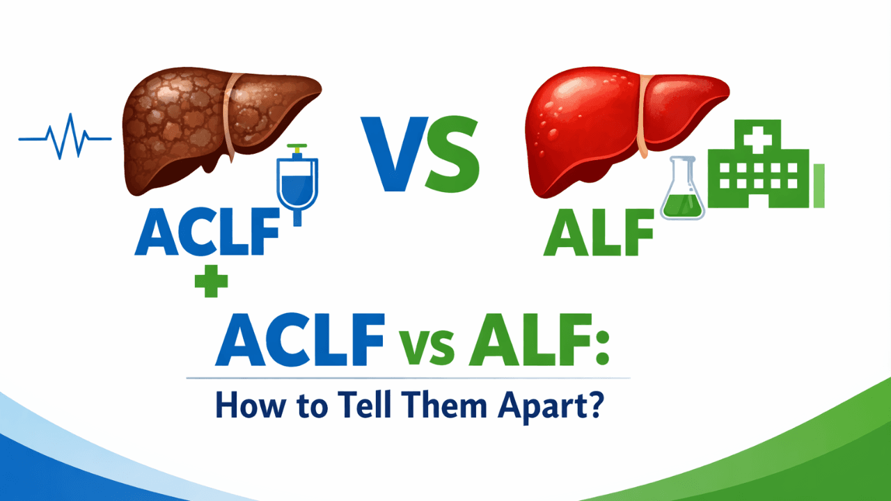
ACLF vs ALF: How to Tell Them Apart and Why It Changes Everything About Treatment
6 min readApr 6, 202624reads

ACLF vs ALF: How to Tell Them Apart and Why It Changes Everything About Treatment

A 58-year-old man with known alcohol-related cirrhosis is admitted with jaundice, confusion, and a bilirubin of 18 mg/dL. Three beds down, a 24-year-old woman with no liver history presents with the same triad — jaundice, encephalopathy, bilirubin of 22 mg/dL — after two weeks of paracetamol overuse. Both patients look critically unwell. Both will be labelled "liver failure" in the initial clerking note. But the diagnosis, prognosis, and management for these two patients are so fundamentally different that conflating them is one of the more consequential errors in hepatology. The distinction between acute-on-chronic liver failure and acute liver failure matters not just academically but at every decision point: who gets admitted to the ICU, who gets listed for transplant, which vasopressors are appropriate, how long you wait before escalating. The challenge is that on a busy admissions ward, the two can look identical for the first few hours. Both present with jaundice, encephalopathy, and coagulopathy. Both can deteriorate within 24 hours. And the underlying liver disease in ACLF may never have been formally diagnosed before. This post is specifically about the diagnostic framework that separates them — and what changes downstream once you get it right. The Defining Difference: Underlying Liver Architecture The single most important question to answer is whether the liver that has failed was previously normal or previously diseased. That question determines everything else. Acute liver failure, as defined by the American Association for the Study of Liver Diseases (AASLD), is the development of coagulopathy (INR ≥1.5) and encephalopathy within 26 weeks in a patient without pre-existing liver disease. The pathophysiology is a sudden, catastrophic hepatocyte loss — from paracetamol toxicity, viral hepatitis A or E, autoimmune hepatitis, Wilson's disease, or ischaemia — in a liver with normal architecture and no fibrosis. The key implication: this liver has full regenerative potential. If the insult is removed and the patient is supported through the acute phase, spontaneous recovery is possible. This is why the transplant decision in ALF is time-limited and reversible — the listing criteria are designed to identify those who will not recover spontaneously, not those who can never regenerate. ACLF is a different beast. Defined by the EASL-CLIF consortium criteria from the landmark CANONIC study, ACLF requires an acute decompensation event — infection, alcohol-related hepatitis, GI bleeding, or an unknown precipitant — superimposed on chronic liver disease, with evidence of organ failure by the EASL-CLIF criteria (renal, hepatic, cerebral, coagulation, circulatory, or respiratory). The underlying liver has already lost architectural integrity. There is no regenerative reservoir. The acute decompensation tips a compensated or previously decompensated cirrhotic into a state of systemic inflammation and multi-organ failure that carries a 28-day mortality of 30–40% in ACLF grade 2, and over 60% in grade 3. Clinical scenario A 47-year-old man presented to the emergency department with a three-day history of increasing jaundice, abdominal distension, and confusion. His family reported he had been drinking heavily for 20 years but had never been formally diagnosed with liver disease. On examination: encephalopathy grade II, bilateral asterixis, tense ascites, spider naevi, palmar erythema. INR 2.8, bilirubin 24 mg/dL, creatinine 2.1 mg/dL, sodium 128 mEq/L. No history of paracetamol use. No recent travel. The clinical picture pointed immediately to ACLF, not ALF — the stigmata of chronic liver disease alone disqualified an ALF diagnosis. The precipitant was identified as an alcohol binge superimposed on compensated cirrhosis. He was graded ACLF-2 by EASL-CLIF criteria (renal and hepatic organ failure). The management priority shifted immediately: not spontaneous recovery, but aggressive precipitant treatment, infection surveillance, and hepatology review for transplant candidacy assessment. He was not listed under ALF criteria — he was evaluated under the ACLF-specific transplant pathway, which has fundamentally different timing and reversibility assumptions. Why the ACLF vs ALF Distinction Drives Completely Different Treatment Decisions Once you have the correct diagnosis, three management decisions diverge immediately. Transplant listing pathway. In ALF, listing is urgent and time-sensitive — King's College Criteria or Clichy criteria guide the decision, and the window for listing can close within 48–72 hours as cerebral oedema develops. In ACLF, the transplant consideration is different: ACLF-3 carries such high 28-day mortality that early transplantation is increasingly supported by evidence, but the patient must first survive the precipitant treatment window. Per the 2023 EASL guidelines on ACLF, patients with ACLF-3 who survive 3–7 days of intensive management should be urgently evaluated for transplant — the listing decision is not immediate but has a defined reassessment window. Precipitant identification and treatment. In ALF, the precipitant is usually known or quickly apparent — paracetamol levels, viral serology, autoimmune panel, Wilson's screen. In ACLF, the precipitant search is broader and the stakes of missing it are higher: 30–40% of ACLF cases are precipitated by bacterial infection, and a significant proportion of those infections are spontaneous bacterial peritonitis with no overt clinical signs. Diagnostic paracentesis is mandatory in any ACLF patient with ascites — not optional, not deferred. Renal management. Hepatorenal syndrome-AKI (HRS-AKI) behaves differently in ACLF versus decompensated cirrhosis without ACLF. Terlipressin response rates are lower, and dose escalation thresholds should be reviewed at 48 hours, not 96 hours. Renal replacement therapy decisions in ACLF-3 need to account for the fact that renal recovery is less likely without transplant. A frequently overlooked point: the "no prior diagnosis" trap in ACLF The most common error in differentiating ACLF from ALF is assuming that a patient cannot have ACLF because they have no prior liver diagnosis. This is wrong, and consequential. In the CANONIC study, a significant proportion of ACLF patients had compensated cirrhosis that had never been clinically identified. The presence of physical stigmata — spider naevi, palmar erythema, gynaecomastia, Dupuytren's contracture, parotid enlargement — combined with a platelet count below 150,000/µL and a low albumin should raise immediate suspicion for underlying chronic disease. A single ultrasound showing a nodular liver surface closes the argument. If there is any doubt, managing the patient as ACLF is the safer default — the consequence of under-treating ACLF as ALF (waiting for spontaneous recovery that cannot come) is far more dangerous than the reverse. Bottom line for clinical practice The first question at any liver failure presentation is not "how bad is it?" — it is "was the liver previously normal?" Stigmata of chronic disease, low platelets, and low albumin answer this before any biopsy or imaging. ACLF grade is determined by the number of organ failures using EASL-CLIF criteria — not by bilirubin level alone. Grade 3 (three or more organ failures) carries >60% 28-day mortality and demands immediate transplant evaluation. In any ACLF patient with ascites, diagnostic paracentesis is mandatory on admission — bacterial infection precipitates 30–40% of ACLF episodes, often silently. Do not apply King's College Criteria to an ACLF patient — they were derived from ALF populations and are not validated for chronic liver disease backgrounds. Terlipressin response in ACLF-related HRS-AKI should be formally reassessed at 48 hours, not the standard 96-hour window used in non-ACLF settings. The next time you admit a patient with jaundice and encephalopathy, GastroAGI can walk you through the ACLF vs ALF differentiation in real time — enter the clinical details and get a structured, guideline-anchored assessment of diagnosis, grade, precipitant checklist, and next steps within seconds. Use GastroAGI For Free.

GastroAGI TeamRead more
Previous
11011
Next
GastroAGI Logo

We are pioneers in clinical intelligence, dedicated to helping gastroenterologists harness the power of artificial intelligence to drive precision, efficiency, and patient growth.

For You

For StudentsFor CliniciansFor ResearchersFor Patients

Core Tools

MELD-Na ScoreChild-PughFIB-4 IndexGlasgow-BlatchfordBISAP Score

Explore

OverviewAboutCalculators
Trending Topics
Conference Briefings
Blog Insights
©GastroAGI 2026
Privacy PolicyTerms of UseMedical Disclaimer