GastroAGI Logo
OverviewBlogsAbout
Trending TopicsDaily BriefConference

Gastroenterology Insights — Clinical Updates, Guides & Resources — Page 5

Left Lateral vs Supine Positioning for EUS-Guided Portal Pressure Measurement: Does Position Matter?
6 min readJul 15, 202619reads

Left Lateral vs Supine Positioning for EUS-Guided Portal Pressure Measurement: Does Position Matter?

Introduction Endoscopic ultrasound-guided portal pressure gradient measurement, or EUS-PPG , is an emerging technique for evaluating portal hypertension. Instead of relying only on indirect assessment, EUS-PPG allows direct pressure measurement by accessing the portal vein and hepatic vein under EUS guidance. A recent review describes EUS-PPG as a modality in which portal pressure is measured by introducing a needle into the hepatic vein and portal vein. Traditionally, portal pressure assessment has been dominated by hepatic venous pressure gradient, or HVPG , which remains the established reference standard for portal hypertension assessment. But as EUS-based hepatology interventions expand, the details of technique matter: patient position, sedation, needle stability, coughing, safety, and measurement reproducibility. A new study in Gastrointestinal Endoscopy asked a practical but important question: Can EUS-PPG be performed reliably in the left lateral position, or does the patient need to be supine? This question matters because routine EUS is commonly performed in the left lateral position, while EUS-PPG has often been performed in the supine position. If left lateral positioning produces comparable PPG values, it could simplify workflow and potentially reduce procedure-related challenges. Why this update matters This study is not about a new drug, device, or guideline. Its importance lies in procedural standardization. EUS-PPG is still a developing technique. For centers that are adopting it, small technical decisions can affect safety, feasibility, training, and reproducibility. Positioning is one of those decisions. A position that improves patient stability, reduces coughing, and avoids repositioning may make the procedure easier and potentially safer. The clinical relevance is especially clear for advanced endoscopists working at the intersection of hepatology and EUS. As EUS-guided liver biopsy, variceal therapy, portal pressure measurement, and other portal hypertension interventions evolve, procedural efficiency and consistency become increasingly important. This study suggests that left lateral positioning may be a reasonable approach for EUS-PPG, at least in the study setting, because portal pressure gradient values were consistent with supine measurements. What the study did This was a prospective single-center study involving patients with portal hypertension undergoing EUS-guided portal pressure gradient measurement under moderate sedation. The investigators measured portal vein pressure and hepatic vein pressure sequentially in both the left lateral and supine positions, without repeat puncture. Portal pressure gradient was calculated as the mean portal vein pressure minus the mean hepatic venous pressure. The study screened 35 patients. Of these, 31 consented, and 29 completed measurements in both positions. In two patients, supine measurements could not be completed because coughing during repositioning dislodged the needle. This detail is clinically relevant. The study was not only testing numerical agreement between two positions. It was also capturing feasibility issues that matter in real-world EUS procedures. What the study found The key finding was that although absolute portal vein pressure and hepatic venous pressure were lower in the left lateral position than in the supine position, the portal pressure gradient itself was similar between the two positions. The reported PPG was 17.2 ± 7.4 mmHg in the supine position and 16.7 ± 6.9 mmHg in the left lateral position , with no statistically significant difference. PPG values showed a strong correlation between positions, with a correlation coefficient of r = 0.978 , and Bland-Altman analysis showed good agreement. The authors also reported fewer coughing events in the left lateral position. Their conclusion was that PPG measurements were consistent between left lateral and supine positioning, and that left lateral positioning under moderate sedation may improve procedural safety and convenience. Clinical interpretation The most important message is that the gradient appears stable even when absolute pressures change . This makes physiologic sense. Portal pressure gradient is a difference between portal venous and hepatic venous pressures. If both pressure readings shift in the same direction with body position, the final gradient may remain clinically similar. For endoscopists, the finding is reassuring. It suggests that EUS-PPG may not require supine positioning in every case. If left lateral positioning gives comparable PPG values, the procedure may align better with standard EUS workflow. The coughing signal is also relevant. Coughing during an EUS-guided vascular puncture is not trivial. Needle stability matters when accessing vascular structures. In this study, coughing during repositioning led to needle dislodgement and prevented completion of supine measurements in two patients. However, this should not be overinterpreted. The study was small, single-center, and performed under moderate sedation. It does not prove that left lateral positioning is universally safer, nor does it define the best position for all sedation strategies, patient anatomies, or disease settings. Practical implications for gastroenterologists For centers already performing EUS-PPG, this study supports a practical workflow question: Can the procedure be done in the left lateral position without compromising the portal pressure gradient result? Based on this study, the answer appears to be yes, with caution. Left lateral positioning may reduce the need for repositioning, fit more naturally into routine EUS practice, and reduce coughing events under moderate sedation. This may be particularly useful in patients who are difficult to reposition, prone to coughing, or undergoing combined EUS procedures. For advanced endoscopy units, the study may support local protocol review. If EUS-PPG is being introduced, positioning should be standardized and documented. Units should track feasibility, adverse events, sedation type, needle dislodgement, technical success, and agreement with clinical findings. For hepatologists, the study is a reminder that portal pressure numbers are not just abstract hemodynamic values. Technique influences measurement. As EUS-PPG becomes more available, clinicians interpreting the results should understand how the measurement was performed. Limitations and caution This study should be viewed as early but useful procedural evidence . The sample size was small. Only 29 patients completed measurements in both positions. The study was conducted at a single center, which may limit generalizability. Operator expertise, sedation practices, equipment, and patient selection may differ across institutions. The study compared positions during the same procedure without repeat puncture, which is methodologically practical but may not fully represent independent measurements on different days or across different operators. It also does not establish whether left lateral positioning should replace supine positioning in all cases. Patients with altered anatomy, severe respiratory compromise, massive ascites, unusual vascular access windows, or deep sedation/general anesthesia may require individualized decisions. Most importantly, this study does not change the clinical thresholds or management of portal hypertension by itself. It is about how to measure , not how to treat . GastroAGI takeaway This Gastrointestinal Endoscopy study addresses a deceptively simple but clinically relevant question: does patient position affect EUS-guided portal pressure gradient measurement? The answer appears reassuring. In this prospective single-center study, left lateral and supine positioning produced consistent PPG values, while left lateral positioning was associated with fewer coughing events under moderate sedation. For advanced endoscopists, the practical implication is that left lateral EUS-PPG may be feasible, convenient, and potentially safer in selected settings. For hepatologists, the message is to pay attention to technique when interpreting portal pressure measurements. This is not yet a practice-changing study. But it is exactly the kind of procedural standardization research that will matter as EUS-guided hepatology continues to mature.

GastroAGI TeamRead more
Alcohol-Associated Hepatitis Is Rising, But the Global Data Gap Remains Large
5 min readJul 15, 202622reads

Alcohol-Associated Hepatitis Is Rising, But the Global Data Gap Remains Large

Introduction Alcohol-associated hepatitis is one of the most severe clinical presentations of alcohol-associated liver disease. It can present acutely with jaundice, systemic inflammation, hepatic decompensation, infection risk, renal dysfunction, and high short-term mortality. Yet despite its clinical severity, the population-level epidemiology of alcohol-associated hepatitis has remained surprisingly difficult to define. A new systematic review published in Clinical Gastroenterology and Hepatology addresses this gap by examining population-based studies reporting the incidence and prevalence of alcohol-associated hepatitis from 2000 to 2025. The review included 11 population-based studies from seven countries and found highly variable incidence estimates, ranging from 1.02 per 100,000 inhabitants in Iceland to 98.5 per 100,000 inhabitants in the United States. The key message is not simply that alcohol-associated hepatitis is increasing. The deeper issue is that our ability to measure its burden remains inconsistent across countries, health systems, coding definitions, and study methods. Why this update matters For gastroenterologists and hepatologists, alcohol-associated hepatitis is not an abstract epidemiologic diagnosis. It is a high-risk clinical syndrome that often enters the healthcare system through emergency departments, inpatient hepatology consults, intensive care units, and transplant evaluation pathways. The new review matters because it highlights a major mismatch: alcohol-associated hepatitis is clinically serious, but the global epidemiologic data remain incomplete. The authors specifically note that reliable epidemiological data are limited despite the high mortality associated with the condition. This matters for several practical reasons. First, health systems need accurate estimates to plan hepatology services, addiction medicine integration, transplant referral capacity, and inpatient care pathways. Second, rising incidence may reflect changing alcohol consumption patterns, delayed presentation, coding differences, pandemic-related alcohol harms, or true increases in disease burden. Third, without standardized case definitions, comparing countries or tracking trends over time becomes difficult. Finally, alcohol-associated hepatitis sits at the intersection of hepatology, addiction medicine, public health, and health policy. Poor epidemiologic visibility can delay prevention and early intervention. What the systematic review found The review searched MEDLINE, Scopus, and the Cochrane Library for population-based studies reporting alcohol-associated hepatitis rates. Risk of bias was assessed using the Joanna Briggs Institute Critical Appraisal Checklist for prevalence studies. The authors included 11 population-based studies from seven countries. Reported annual incidence rates varied widely, from 1.02 per 100,000 inhabitants in Iceland to 98.5 per 100,000 inhabitants in the United States. The median incidence rate was 6.8 cases per 100,000 inhabitants, with an interquartile range of 4.2–19. Among nine studies that reported at least two estimates over time, seven showed increasing incidence, while two showed a decrease. A sensitivity analysis excluding studies affected by the COVID-19 pandemic still showed a similar pattern: incidence increased in five of seven studies. The pandemic signal is also important. Two studies reported a surge in alcohol-associated hepatitis incidence during the COVID-19 pandemic. One study with follow-up beyond the pandemic observed a gradual return toward pre-pandemic levels. One of the most striking findings was the absence of prevalence data. The review found no studies reporting population-based prevalence of alcohol-associated hepatitis. Clinical interpretation The review does not change how gastroenterologists diagnose or treat alcohol-associated hepatitis at the bedside. It does not provide a new treatment algorithm, prognostic score, or therapeutic recommendation. Its value lies elsewhere: it shows that the burden of alcohol-associated hepatitis is probably under-characterized and inconsistently measured. The wide variation in incidence estimates should not be interpreted as purely biological or geographic variation. Some of the difference likely reflects how alcohol-associated hepatitis is defined, coded, captured, and studied. For example, administrative coding may miss milder cases, misclassify decompensated alcohol-associated cirrhosis, or vary across healthcare systems. At the same time, the overall trend toward increasing incidence across most longitudinal studies is clinically concerning. It suggests that alcohol-associated hepatitis may be becoming more common, or at least more frequently recognized and recorded. The COVID-19 findings should be interpreted cautiously. Pandemic-related increases in alcohol use, delayed healthcare access, psychosocial stress, and healthcare system disruption may all have contributed. However, one study showing a gradual return toward pre-pandemic levels suggests that not all pandemic-era surges necessarily represent a permanent new baseline. Practical implications for gastroenterologists For hepatology practice, this review reinforces the need to view alcohol-associated hepatitis as both an acute clinical emergency and a population-health problem. At the patient level, clinicians should continue to focus on early recognition, severity stratification, infection assessment, renal monitoring, nutrition, alcohol cessation support, addiction medicine involvement, and appropriate transplant referral discussions. At the system level, the review supports better registry design, standardized diagnostic definitions, and collaboration between hepatology, public health, and addiction services. For trainees, this paper is a reminder that epidemiology is not just background data. In alcohol-associated hepatitis, incidence trends affect workforce planning, inpatient hepatology demand, ICU utilization, transplant ethics, and prevention strategy. For researchers, the absence of population-level prevalence data is a clear gap. Future studies need harmonized case definitions, prospective designs, and better differentiation between alcohol-associated hepatitis, alcohol-associated cirrhosis with decompensation, and other causes of acute-on-chronic liver failure. Limitations and caution This is a systematic review of population-based studies, so its conclusions depend on the quality and consistency of the underlying studies. The authors found major heterogeneity across estimates, likely due to differences in case definitions, methodology, coding practices, healthcare systems, and true geographic variation. The review should not be used to claim a precise global incidence of alcohol-associated hepatitis. Instead, it supports a more cautious conclusion: available population-based studies suggest an increasing trend, but the true global burden remains poorly defined. It is also important not to overstate practice implications. This paper does not prove that any specific intervention reduces alcohol-associated hepatitis incidence. It does, however, strengthen the rationale for better surveillance, earlier alcohol-use interventions, and integrated hepatology-addiction care models. GastroAGI takeaway Alcohol-associated hepatitis remains a severe and high-mortality manifestation of alcohol-associated liver disease, but its global epidemiology is still not well measured. This systematic review suggests that incidence has increased in many population-based studies over the past 25 years, with additional pandemic-era surges reported in some settings. For gastroenterologists, the message is clear: alcohol-associated hepatitis deserves attention not only as an inpatient syndrome, but also as a growing public health and systems-of-care challenge. Better epidemiology will not treat the patient in front of us today. But without better epidemiology, we cannot accurately plan services, allocate resources, build prevention strategies, or understand whether the burden is truly rising. Reference/source Clinical Gastroenterology and Hepatology , “Epidemiology of Alcohol-Associated Hepatitis: A Systematic Review of Population-Based Studies,” published online July 2026.

GastroAGI TeamRead more
BSG 2026 Adult Coeliac Disease Guideline: What Gastroenterologists Need to Know
6 min readJul 15, 202641reads

BSG 2026 Adult Coeliac Disease Guideline: What Gastroenterologists Need to Know

Introduction The British Society of Gastroenterology has released its 2026 guideline on the diagnosis and management of adult coeliac disease , updating standards for a condition that remains common, under-recognised, and clinically heterogeneous. The guideline was developed by a multidisciplinary panel and focuses on evidence-based diagnosis, long-term management, follow-up, nutritional assessment, and refractory coeliac disease care. For gastroenterologists, this update is important because coeliac disease is no longer just a “positive serology plus duodenal biopsy” diagnosis in every adult. The guideline acknowledges newer evidence around serology-based diagnosis, gluten challenge, follow-up intensity, mucosal healing, bone health, vaccination, and referral pathways for complex disease. The practical message is not that biopsy has become obsolete. Rather, the guideline moves adult coeliac care toward a more individualised model: test correctly, biopsy when needed, use no-biopsy diagnosis only in selected circumstances, support the gluten-free diet properly, and identify patients who need structured long-term review. Why this update matters Coeliac disease affects the small intestine but often presents beyond the gut. Patients may present with diarrhoea, bloating, weight loss, iron deficiency anaemia, osteoporosis, abnormal liver enzymes, infertility, neurological symptoms, or fatigue. Others are diagnosed through risk-based testing. The BSG guideline notes that coeliac disease is a chronic autoimmune disorder in genetically predisposed individuals, with a reported prevalence of approximately 1%. It also reiterates that a lifelong gluten-free diet remains the only accepted treatment. The update matters clinically for three reasons. First, diagnostic pathways are evolving. Adults with strongly positive IgA-tTG may, in selected circumstances, be diagnosed without duodenal biopsy, but this requires careful selection and shared decision-making. Second, follow-up is being reframed. Rather than assuming every patient needs indefinite routine clinic review, the guideline supports structured follow-up early after diagnosis, then patient-initiated follow-up for stable responders, while reserving systematic long-term follow-up for higher-risk groups. Third, the guideline gives practical direction for non-responsive and refractory coeliac disease, including when to refer to specialist centres and how to assess suspected refractory coeliac disease. What the guideline found A key diagnostic recommendation is that patients should be consuming a gluten-containing diet before testing. The guideline recommends IgA-tissue transglutaminase antibodies as the first-line investigation and advises screening for selective IgA deficiency alongside IgA-tTG testing. When biopsy is required, the guideline recommends taking at least four biopsies from the second part of the duodenum and two from the duodenal bulb . This is clinically important because patchy disease and inadequate sampling can lead to missed diagnosis. One of the most discussed updates is the optional adult no-biopsy pathway. In symptomatic adults assessed in secondary care, the guideline suggests that coeliac disease can be diagnosed without duodenal biopsies when IgA-tTG is at least 10 times the upper limit of normal . However, this is a conditional recommendation, intended for shared decision-making rather than automatic use. The guideline also cautions against using a lower tTG threshold for a no-biopsy diagnosis because it may increase false-positive diagnoses. It also advises caution in patients with recent autoimmune diagnoses, where false elevation of tTG may be a concern. For patients already on a gluten-free diet before proper testing, the guideline recommends a gluten challenge of 3–6 g of gluten daily for at least 6 weeks . It notes that duodenal histology is preferred over serology as the diagnostic readout because seroconversion after 6 weeks may be low. Management continues to centre on a lifelong gluten-free diet. The guideline recommends that patients diagnosed with coeliac disease maintain a lifelong gluten-free diet to improve symptoms, quality of life, and reduce complications. It also recommends a combined approach to assessing gluten-free diet adherence because no single test is adequate alone. Follow-up is another major practical area. The guideline advises regular follow-up for up to 2 years after diagnosis. After that, patients who respond well may be considered for patient-initiated follow-up, while those with poor adherence, inadequate response, or complications should receive longer-term systematic follow-up. Nutritional monitoring is emphasised. The guideline advises assessment and monitoring of iron, folate, vitamin B12, vitamin D, and calcium at diagnosis and during follow-up. It also advises DXA scanning 1 year after starting a gluten-free diet in newly diagnosed adults. For refractory disease, the guideline recommends referral to a specialist centre with an experienced multidisciplinary team. It also recommends flow cytometry to immunophenotype small intestinal intraepithelial lymphocytes as the reference standard for diagnosing refractory coeliac disease type 2. Clinical interpretation The most clinically relevant shift is the guideline’s balanced position on the no-biopsy pathway. It does not remove the role of endoscopy. Instead, it defines where biopsy may be avoided and where it remains essential. This distinction matters. A patient with classic symptoms, high IgA-tTG, appropriate clinical setting, and informed preference may reasonably avoid biopsy under the guideline framework. But patients with equivocal serology, IgA deficiency, iron deficiency anaemia undergoing endoscopy, possible seronegative disease, alarm features, or concern for alternative diagnoses still need careful endoscopic and histological assessment. The gluten challenge recommendation is also practical. Many patients start a gluten-free diet before seeing a gastroenterologist. Without adequate gluten exposure, both serology and histology may become falsely reassuring. The guideline’s 3–6 g daily for at least 6 weeks gives clinicians a usable framework, while still recognising tolerability issues. The follow-up model is also realistic. Clinics cannot provide indefinite specialist follow-up for every stable patient, but early follow-up is important for diet education, symptom assessment, nutritional correction, and adherence support. Patient-initiated follow-up may be reasonable for stable responders, but not for patients with persistent symptoms, abnormal labs, poor adherence, persisting villous atrophy, or complications. Practical implications for gastroenterologists For daily practice, the guideline supports a few clear changes. Do not test for coeliac disease after gluten has already been removed unless the limitations are clearly understood. Encourage gluten-containing diet before initial testing where clinically safe. Use IgA-tTG first line, but remember to assess IgA deficiency. In selective IgA deficiency, IgG-based serology and duodenal biopsy become more important. When performing endoscopy for suspected coeliac disease, take enough biopsies: four from D2 and two from the bulb. Under-sampling remains a preventable cause of diagnostic uncertainty. Consider no-biopsy diagnosis only in selected symptomatic adults in secondary care with IgA-tTG ≥10× upper limit of normal, and only after shared decision-making. For patients already gluten-free, discuss a structured gluten challenge rather than relying on negative tests. Follow patients actively for the first 2 years after diagnosis, then individualise long-term review. Assess micronutrients and bone health, and remember that persistent symptoms after a gluten-free diet require a systematic approach rather than simply advising “stricter gluten avoidance.” Limitations and caution This is a guideline, not a single new trial. Some recommendations are based on high or moderate certainty evidence, while others are conditional, consensus-based, or expert opinion. The no-biopsy pathway should not be applied casually, and it may not suit patients with atypical features, possible alternative diagnoses, or situations where histology would change management. The guideline also acknowledges that follow-up decisions may need to be adapted to local healthcare resources. Patient-initiated follow-up is not the same as discharge without safety-netting. GastroAGI takeaway The 2026 BSG adult coeliac disease guideline is a clinically useful update for gastroenterologists. Its main message is precision: diagnose on a gluten-containing diet, use IgA-tTG correctly, biopsy properly when needed, reserve no-biopsy diagnosis for carefully selected adults, support the gluten-free diet with structured follow-up, and refer suspected refractory disease early. For clinicians, this is a practical roadmap for moving adult coeliac disease care from a one-size-fits-all model toward a more accurate, patient-centred, and risk-based approach. Reference/source British Society of Gastroenterology guideline page and full guideline PDF.

GastroAGI TeamRead more
Sphingolipid Metabolism and KRAS in Pancreatic Cancer: A New Translational Signal
7 min readJul 13, 202619reads

Sphingolipid Metabolism and KRAS in Pancreatic Cancer: A New Translational Signal

Introduction Pancreatic ductal adenocarcinoma remains one of the most difficult cancers in GI oncology. Despite progress in systemic therapy, surgery, molecular profiling, and supportive care, pancreatic cancer continues to be defined by late diagnosis, aggressive biology, early metastasis, and limited durable treatment responses. One of the central biological drivers of pancreatic cancer is KRAS signaling. KRAS-mutant biology has long been recognized as fundamental to pancreatic carcinogenesis, but targeting KRAS directly has historically been challenging. The emergence of mutant-selective KRAS inhibitors has renewed interest in understanding what allows KRAS-driven tumors to survive, signal, adapt, and resist therapy. A new Gut study adds an important mechanistic layer to this discussion. The article, titled “Dysregulated sphingolipid metabolism drives pancreatic carcinogenesis through plasma membrane Kras enrichment,” explores how altered sphingolipid metabolism may influence KRAS localization and oncogenic signaling in pancreatic ductal adenocarcinoma. The study focuses on SMPD1 , also known as acid sphingomyelinase, an enzyme involved in sphingolipid metabolism. SMPD1 converts sphingomyelin to ceramide, a lipid involved in membrane structure and signaling regulation. For clinicians, the key point is not that SMPD1 is ready to become a therapeutic target tomorrow. The more useful takeaway is that pancreatic cancer biology may depend not only on genetic drivers such as KRAS, but also on the lipid environment that helps organize oncogenic signaling at the cell membrane. Why this update matters The clinical relevance of this study comes from the intersection of three major themes in pancreatic cancer research: KRAS dependency, metabolic rewiring, and treatment resistance. KRAS signaling depends on proper cellular localization. It is not enough for a cancer cell to carry an oncogenic KRAS mutation; KRAS must also interact with the right cellular membrane compartments to transmit downstream signals. This study suggests that sphingolipid metabolism, through SMPD1-dependent changes, may support KRAS enrichment at the plasma membrane and thereby strengthen oncogenic signaling. That is clinically interesting because it reframes lipid metabolism as more than a background metabolic feature. It may actively shape how oncogenic signaling is organized. The study also matters because pancreatic cancer therapy is moving toward more targeted approaches. If KRAS signaling can be influenced by the lipid composition of the plasma membrane, then metabolic pathways may eventually become rational combination partners with KRAS-directed therapies. However, this remains a research concept, not a clinical recommendation. What the study found According to the study summary available through PubMed and Gut , tumor cell-autonomous expression of SMPD1 in pancreatic ductal adenocarcinoma was associated with poorer patient outcomes. In experimental models, Smpd1 ablation in murine PDAC cells reduced proliferation and migration in vitro and decreased metastases and tumor burden in vivo. The investigators used integrated transcriptomic, metabolomic, and proteomic analyses. These approaches suggested that disrupting SMPD1 impaired KrasG12D oncogenic signaling , which was linked to reduced tumor burden. Mechanistically, reduced plasma membrane interaction of KrasG12D was associated with SMPD1-dependent sphingolipid metabolism. In simpler terms, altered sphingolipid metabolism appeared to influence whether oncogenic KRAS was enriched at the plasma membrane, where it can effectively signal. The study also reported that inhibition of Smpd1 was synergistic with KrasG12D inhibition , which is one of the most clinically intriguing parts of the work. This does not mean that SMPD1 inhibition is ready for pancreatic cancer treatment. It does suggest that membrane lipid biology may become an important part of future KRAS-targeted combination strategies. Clinical interpretation For gastroenterologists, hepatopancreatobiliary specialists, GI oncologists, and researchers, this study should be read as a mechanistic signal rather than a clinical directive. The major clinical idea is that pancreatic cancer may use lipid metabolism to support oncogenic signaling architecture. KRAS activity is not only determined by mutation status. It is also shaped by cellular context, membrane localization, metabolic dependencies, feedback pathways, and tumor microenvironment interactions. SMPD1 is especially interesting because it connects metabolism and membrane biology. Acid sphingomyelinase converts sphingomyelin into ceramide, and this process can alter membrane properties and signal transduction. If SMPD1 helps organize a membrane environment that favors KRAS signaling, then blocking this pathway could theoretically weaken KRAS-driven tumor behavior. The study’s finding of synergy between Smpd1 inhibition and KrasG12D inhibition makes this hypothesis more compelling, but it remains preclinical. The clinically responsible interpretation is this: This study identifies a potentially important KRAS-supporting mechanism in pancreatic cancer, but it does not yet establish a new treatment pathway. Practical implications for gastroenterologists and GI oncology teams For clinicians, this study has no immediate effect on standard pancreatic cancer management. Patients should continue to be treated according to established multidisciplinary pathways, including staging, surgical assessment, systemic therapy, radiation where appropriate, molecular profiling when indicated, nutritional support, pain control, biliary or gastric outlet obstruction management, and clinical trial consideration. The main practical value is educational and research-oriented. First, it provides a useful way to explain why pancreatic cancer remains biologically difficult. KRAS mutation status is important, but KRAS-driven tumors are supported by complex cellular systems, including lipid metabolism and membrane organization. Second, it reinforces the value of translational research. Studies like this help identify vulnerabilities that may not be obvious from sequencing alone. A tumor may carry a KRAS mutation, but its dependence on membrane lipid biology could influence how strongly that pathway signals or how it responds to targeted inhibition. Third, it supports the rationale for combination strategies. Future pancreatic cancer trials may increasingly combine direct oncogene inhibition with agents that alter signaling dependencies, metabolic state, tumor immune contexture, stromal interactions, or adaptive resistance pathways. Fourth, it highlights why clinicians should be cautious when communicating early science. This is not evidence that a supplement, lipid-lowering strategy, dietary manipulation, or off-label SMPD1-targeting approach treats pancreatic cancer. The study is mechanistic and should remain in the research domain until clinical evidence emerges. Limitations and caution The most important limitation is that this is preclinical and translational work. The study includes experimental models and associations with patient outcomes, but it does not test an SMPD1-targeting therapy in pancreatic cancer patients. Experimental ablation or inhibition in models does not automatically translate into safe or effective treatment in humans. Pancreatic cancer is highly heterogeneous, and tumor metabolism can vary across patients, disease stages, treatment exposures, and microenvironmental contexts. Another caution is that lipid metabolism is complex. Ceramide and sphingolipid pathways can have different effects depending on cell type, subcellular compartment, timing, and disease context. A pathway that appears tumor-promoting in one setting may not behave identically in another. The synergy with KrasG12D inhibition is scientifically important, but it should be interpreted as a rationale for further research, not as a treatment recommendation. GastroAGI takeaway This Gut study provides a compelling translational insight: dysregulated sphingolipid metabolism, through SMPD1, may help pancreatic cancer cells enrich oncogenic KRAS at the plasma membrane and strengthen tumor-promoting signaling. For clinicians, the message is not to change practice today. The message is to understand where pancreatic cancer research is moving. KRAS-targeted therapy is no longer only about blocking the mutant protein. It may also require understanding the cellular environment that allows KRAS to signal effectively. SMPD1 and sphingolipid metabolism may represent one such supportive mechanism. The GastroAGI angle is clear: In pancreatic cancer, the next generation of KRAS strategies may need to look beyond the mutation itself—and into the membrane biology that helps KRAS drive disease. Key Points A new Gut study links dysregulated sphingolipid metabolism to pancreatic carcinogenesis through plasma membrane Kras enrichment. The study focuses on SMPD1 , an acid sphingomyelinase involved in converting sphingomyelin to ceramide. Tumor cell-autonomous SMPD1 expression in PDAC was associated with poorer patient outcomes. Smpd1 ablation reduced proliferation and migration in vitro and decreased metastases and tumor burden in vivo. Integrated omics suggested that SMPD1 disruption impaired KrasG12D oncogenic signaling. Smpd1 inhibition appeared synergistic with KrasG12D inhibition in experimental models. This is a promising translational signal, but it is not practice-changing and should not guide treatment outside research settings. Reference / Source Alnatsha A, et al. Dysregulated sphingolipid metabolism drives pancreatic carcinogenesis through plasma membrane Kras enrichment. Gut . Published online July 3, 2026. DOI: 10.1136/gutjnl-2025-337363 . Primary source link: Gut Online First article. PubMed record: PubMed indexed abstract and study summary. Optional supporting background reference, only if you want an extra mechanistic citation: Liu J, et al. Glycolysis regulates KRAS plasma membrane localization and function through glycosphingolipid synthesis. Nature Communications . 2023.

GastroAGI TeamRead more
Risk-Based Follow-Up After HCC Ablation: Toward Smarter Surveillance After Curative Therapy
6 min readJul 10, 202626reads

Risk-Based Follow-Up After HCC Ablation: Toward Smarter Surveillance After Curative Therapy

Introduction Hepatocellular carcinoma, or HCC, remains one of the most recurrence-prone cancers managed by hepatologists, gastroenterologists, interventional radiologists, surgeons, and oncology teams. For selected patients with early-stage HCC, local ablation can be a curative-intent therapy. However, the clinical challenge does not end once complete ablation is achieved. The next question is just as important: how should these patients be followed after treatment? A new JHEP Reports article titled “A risk-based post ablation follow-up strategy for hepatocellular carcinoma” addresses this question. The article was available online on July 6, 2026, as an in-press journal pre-proof. The study is clinically relevant because many HCC follow-up pathways still rely on fixed imaging intervals. The article notes that after ablation, patients were recommended routine follow-up at intervals of 3–6 months during the first 2 years and every 6–12 months thereafter. That approach is simple, but it may not be optimal for everyone. Why this update matters HCC recurrence risk after curative-intent therapy is not uniform. Some patients recur early, some recur late, and some remain recurrence-free for long periods. A fixed follow-up schedule may therefore overtreat lower-risk patients while under-serving patients with higher recurrence risk. This matters for several reasons. First, earlier detection of recurrence can preserve the chance of repeat curative-intent therapy, including repeat ablation, resection, transplantation evaluation, or locoregional therapy depending on disease pattern and liver reserve. Second, repeated CT or MRI surveillance is resource-intensive. It adds cost, patient anxiety, contrast exposure, travel burden, and clinic load. Third, HCC care is increasingly moving toward individualized decision-making. We already personalize treatment based on tumor stage, liver function, performance status, portal hypertension, transplant eligibility, and multidisciplinary review. Follow-up after ablation should logically move in the same direction. AASLD maintains HCC practice guidance resources focused on prevention, diagnosis, and treatment, underscoring that HCC care requires evidence-based, multidisciplinary decision-making. What the study examined According to the indexed article information, the investigators retrospectively analyzed patients who received complete ablation for initial HCC within the Milan criteria from two tertiary hospitals in China. The key concept was to move beyond a single fixed schedule and develop a risk-based post-ablation surveillance strategy. In practical terms, this means follow-up intensity could be aligned with estimated recurrence risk rather than applied equally to every patient. This type of study is important because post-treatment HCC surveillance has to balance two competing goals: Detect recurrence early enough to act. Avoid unnecessary imaging and clinic visits in patients whose recurrence risk is relatively lower. The available source information does not support treating this as a definitive guideline. It is better understood as a risk-stratification and surveillance-optimization study. Clinical interpretation The main clinical message is not that current follow-up schedules are wrong. Rather, this study highlights that fixed surveillance intervals may be too blunt for a disease as biologically heterogeneous as HCC. After ablation, recurrence risk may be influenced by tumor-related factors, liver-related factors, treatment-related factors, and host-related factors. Clinicians commonly consider tumor size, tumor number, AFP, liver function, viral hepatitis status, cirrhosis severity, ablation margin, imaging response, and prior recurrence history when estimating risk. A risk-based follow-up model could help identify patients who may benefit from closer surveillance early after ablation, when recurrence risk is concentrated. Conversely, patients with lower predicted risk might avoid unnecessary high-frequency imaging, provided safety is maintained. This is especially relevant in systems where MRI access is limited, CT capacity is strained, or patients travel long distances for surveillance. The most useful way to frame this update for clinicians is: HCC surveillance after ablation may eventually become adaptive, not one-size-fits-all. Practical implications for gastroenterologists and hepatologists For clinicians managing HCC patients after ablation, this article supports several practical reflections. First, post-ablation follow-up should begin with confirmation of complete response. Imaging quality and interpretation matter. If the ablation zone is difficult to assess, or if margins are uncertain, the patient may need closer early reassessment. Second, risk documentation should become routine. Instead of simply writing “follow up in 3 months,” clinicians can document why a patient is considered high, intermediate, or lower risk for recurrence. Third, surveillance planning should be multidisciplinary. Hepatologists, gastroenterologists, radiologists, interventional radiologists, surgeons, oncologists, and transplant teams may each interpret recurrence risk differently. A shared pathway can reduce variation. Fourth, risk-based strategies should not reduce vigilance inappropriately. HCC patients often have cirrhosis or chronic liver disease, meaning they remain at risk not only for local recurrence but also for new intrahepatic tumors. Fifth, patient communication matters. Patients should understand that “less frequent imaging” does not mean less serious care, and “more frequent imaging” does not necessarily mean recurrence is expected. It means surveillance is being matched to risk. Limitations and caution This update should be interpreted carefully. The study is retrospective, which means it can identify patterns and build risk-based strategies, but it cannot prove that a new surveillance schedule improves survival compared with standard care. The reported study population included patients treated at two tertiary hospitals in China and patients with initial HCC within the Milan criteria. That may limit generalizability to other populations, including patients with non-viral liver disease, different transplant pathways, different imaging access, or different ablation techniques. Another important limitation is implementation. A risk-based follow-up model is only useful if it is easy to apply, externally validated, clinically interpretable, and safe across diverse settings. Finally, this should not be viewed as permission to relax follow-up after HCC ablation. Recurrence remains clinically important, and surveillance decisions should still follow local guidelines, tumor board recommendations, liver function assessment, and patient-specific risk. GastroAGI takeaway This JHEP Reports study is a timely reminder that HCC follow-up after ablation may need to become more personalized. The concept is simple but clinically powerful: patients with different recurrence risks may not need identical surveillance schedules. For now, this is best viewed as hypothesis-generating and practice-informing, not practice-changing. But it points toward a future in which post-ablation HCC surveillance is guided by recurrence risk, resource stewardship, and patient-centered follow-up rather than fixed intervals alone. The GastroAGI angle is clear: After curative-intent HCC ablation, the next frontier may be not just better treatment—but smarter surveillance. Key Points A new JHEP Reports article proposes a risk-based follow-up strategy after HCC ablation. The study focused on patients who received complete ablation for initial HCC within the Milan criteria. Routine follow-up after ablation is often recommended every 3–6 months in the first 2 years and every 6–12 months thereafter. A fixed schedule may not match the true recurrence risk of every patient. Risk-based surveillance could help intensify follow-up for higher-risk patients and reduce unnecessary visits for lower-risk patients. The evidence is retrospective and should not be treated as a new guideline yet. The strongest GastroAGI angle: HCC surveillance after ablation may be moving from fixed intervals to individualized risk-based care. Explore GastroAGI If you are a gastroenterologist, trainee, researcher, or medical educator navigating journals, guidelines, conferences, and clinical questions every day, GastroAGI is built for you. GastroAGI brings GI-focused intelligence into one structured platform — helping users learn, reason, and communicate with more clarity across Student, Clinician, and Patient modes. Explore how specialized AI can support modern gastroenterology workflows. Visit GastroAGI: https://gastroagi.com

GastroAGI TeamRead more
VTE After Endoscopic Sleeve Gastroplasty: Rare Event, Important Safety Signal
6 min readJul 10, 202628reads

VTE After Endoscopic Sleeve Gastroplasty: Rare Event, Important Safety Signal

Introduction Endoscopic sleeve gastroplasty, or ESG, is increasingly becoming part of the therapeutic landscape for patients with obesity. It offers a less invasive, incisionless option compared with surgical bariatric procedures and is now moving further into mainstream metabolic endoscopy practice. A new article in Gastrointestinal Endoscopy addresses an important safety question: how often does venous thromboembolism occur after ESG, and what are current prophylaxis patterns? The study, titled “Venous Thromboembolism Following Endoscopic Sleeve Gastroplasty,” was authored by Benjamin M. Moy, Rolando Barajas, Chloe Savino, and Allison R. Schulman. It was published online ahead of print in Gastrointestinal Endoscopy , with DOI 10.1016/j.gie.2026.06.065 . This is clinically relevant because ESG is expanding at the same time that obesity medicine, bariatric endoscopy, and metabolic care pathways are rapidly evolving. As more patients undergo ESG, endoscopists need practical data on uncommon but potentially serious adverse events. Why this update matters Venous thromboembolism, or VTE, includes deep venous thrombosis and pulmonary embolism. Even when uncommon, VTE matters because PE can be life-threatening and because obesity itself is a known risk context for thrombosis. The study is timely because the authors note that there are currently no established guidelines specifically addressing peri-procedural VTE management after ESG. That gap is important. ESG sits between traditional endoscopy and bariatric surgery. It is performed endoscopically, but patients often have metabolic risk factors, elevated BMI, obstructive sleep apnea, limited mobility, prior VTE history, or other comorbidities that may influence peri-procedural risk. The broader field is also changing. The joint ASGE–ESGE guideline provides evidence-based recommendations on endoscopic bariatric and metabolic therapies in adults with obesity, and ESG has also gained increasing recognition in clinical and coding pathways. That makes safety data increasingly important. Clinicians need to know whether VTE after ESG is common enough to justify routine prophylaxis, or rare enough that prophylaxis should remain individualized. What the study found The study analyzed patients who underwent ESG from 2020 to 2023 using the Metabolic and Bariatric Surgery Accreditation and Quality Improvement Program, or MBSAQIP, database. The primary outcome was 30-day post-ESG DVT and PE. Secondary outcomes included VTE prophylaxis methods and comorbid predictors. A total of 2,371 ESG patients were included. The cohort had a mean age of 45 years, was 85.8% female, and had a mean BMI of 40 kg/m². Before ESG, 29 patients, or 1.2%, had a history of DVT, and 38 patients, or 1.6%, were on therapeutic anticoagulation. Prophylaxis patterns varied substantially. Mechanical prophylaxis alone was used in 46.7%, pharmacologic prophylaxis alone in 0.7%, both mechanical and pharmacologic prophylaxis in 12.4%, and no prophylaxis in 40.2%. The 30-day incidence of postoperative DVT was 0.3%, and the incidence of PE was 0.2%. Two-thirds of patients who developed VTE were not receiving prophylaxis. The authors’ conclusion is reassuring but not dismissive: VTE after ESG appears to be extremely low in this large multicenter database, with events occurring in fewer than 0.5% of cases. Clinical interpretation The headline is simple: VTE after ESG is rare. But the clinical interpretation should be more nuanced. Rare does not mean irrelevant, especially when an adverse event can be serious. The key message is not that every ESG patient needs pharmacologic prophylaxis. The better message is that ESG programs should have a clear, risk-based approach to VTE assessment. This study supports the overall safety profile of ESG, but it also shows variation in prophylaxis practice. Nearly half of patients received mechanical prophylaxis alone, while about 40% received no prophylaxis. Pharmacologic-only prophylaxis was uncommon, and combined prophylaxis was used in a minority of patients. That variation likely reflects real-world uncertainty. ESG is not identical to laparoscopic bariatric surgery, but it is also not a routine diagnostic endoscopy. It involves anesthesia, procedural time, obesity-related risk factors, and a post-procedure recovery period. Therefore, a one-size-fits-all approach may be inappropriate. For low-risk patients, the very low VTE rate may argue against routine pharmacologic prophylaxis. For higher-risk patients, including those with prior VTE, marked obesity, limited mobility, thrombophilia, active cancer, estrogen exposure, prolonged procedure time, or other risk factors, more deliberate prophylaxis planning may be reasonable. Practical implications for gastroenterologists For endoscopists and bariatric endoscopy programs, this study should encourage structured pre-procedure planning. First, VTE risk should be assessed before ESG. This does not necessarily require a complex protocol for every patient, but history of prior DVT or PE, anticoagulant use, BMI, mobility, smoking, hormone therapy, thrombophilia, cancer history, and obstructive sleep apnea should be reviewed. Second, prophylaxis decisions should be documented. If mechanical prophylaxis is used, that should be clear. If pharmacologic prophylaxis is withheld, the rationale should be clear, especially in patients with risk factors. Third, anticoagulation management should be individualized. Some patients undergoing ESG may already be on therapeutic anticoagulation. In those cases, clinicians must balance thrombotic risk against bleeding risk from the procedure. Fourth, recovery instructions should include VTE awareness. Patients should know to seek urgent care for unilateral leg swelling, calf pain, sudden shortness of breath, chest pain, syncope, or unexplained tachycardia. This is especially important because many events occur after discharge. Fifth, ESG programs should consider whether their existing bariatric surgery pathways, anesthesia pathways, and endoscopy pathways are aligned. ESG lives at the intersection of all three. Limitations and caution This study is useful, but it should not be overinterpreted. It is an observational database analysis, not a randomized trial of prophylaxis strategies. Therefore, it cannot prove that one prophylaxis approach is superior to another. Patients who received prophylaxis may have had higher baseline risk, and patients who received no prophylaxis may have been lower risk. The study also focuses on 30-day outcomes. Events outside that window would not be captured in the primary outcome. In addition, database studies depend on accurate coding and reporting of events. Most importantly, the finding that VTE incidence is low should not be translated into “no prophylaxis is needed.” Instead, it should support rational risk stratification and avoidance of unnecessary anticoagulation in low-risk patients while maintaining vigilance in higher-risk groups. GastroAGI takeaway VTE after endoscopic sleeve gastroplasty appears to be rare, with reported 30-day DVT and PE rates of 0.3% and 0.2%, respectively. For clinicians, the key point is not to overreact or ignore the signal. ESG remains a generally safe endoscopic bariatric procedure, but expanding procedural volume requires mature safety pathways. The practical GastroAGI message: ESG programs should move toward risk-based VTE assessment, clear prophylaxis documentation, and patient education—rather than routine anticoagulation for everyone or no structured assessment at all. Reference / Source Moy BM, Barajas R, Savino C, Schulman AR. Venous Thromboembolism Following Endoscopic Sleeve Gastroplasty. Gastrointestinal Endoscopy . Published online ahead of print, 2026. DOI: 10.1016/j.gie.2026.06.065. Key Points VTE after ESG appears uncommon in a large multicenter database. The study included 2,371 ESG patients from 2020–2023. Reported 30-day DVT incidence was 0.3%, and PE incidence was 0.2%. Prophylaxis practice varied: mechanical only, pharmacologic only, combined prophylaxis, and no prophylaxis were all represented. Two-thirds of patients who developed VTE were not on prophylaxis. The study supports risk-based assessment, not automatic anticoagulation for all ESG patients. This is awareness-generating and clinically useful, but not by itself practice-changing. Explore GastroAGI If you are a gastroenterologist, trainee, researcher, or medical educator navigating journals, guidelines, conferences, and clinical questions every day, GastroAGI is built for you. GastroAGI brings GI-focused intelligence into one structured platform — helping users learn, reason, and communicate with more clarity across Student, Clinician, and Patient modes. Explore how specialized AI can support modern gastroenterology workflows. Visit GastroAGI: https://gastroagi.com

GastroAGI TeamRead more
MELD-Na Score: How to Interpret It and When It Changes the Transplant Conversation
7 min readJul 9, 20261.2kreads

MELD-Na Score: How to Interpret It and When It Changes the Transplant Conversation

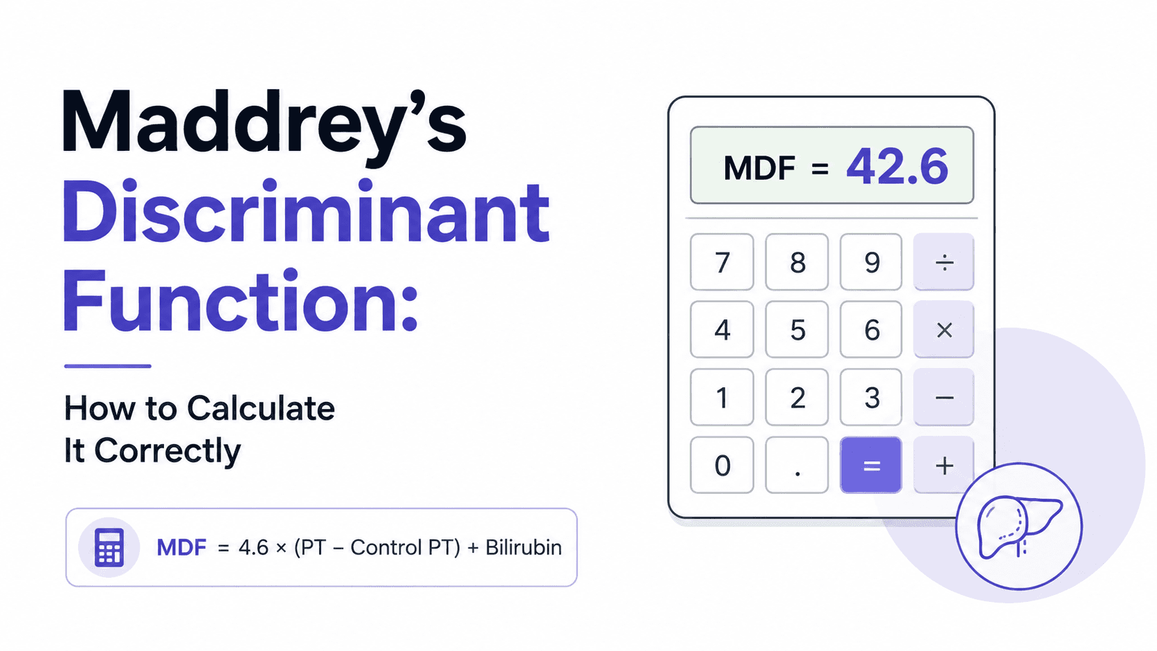
A 58-year-old woman with alcohol-related cirrhosis is admitted for worsening ascites. Her creatinine and INR are stable compared to three months ago, and on a quick glance, her chart looks unremarkable. Her sodium, though, has dropped from 135 to 128 mEq/L over that same window. Her MELD score barely moved. Her MELD-Na score jumped six points. The transplant team's tone in the room changed before the labs were even fully explained to the family - and that shift, the gap between what MELD says and what MELD-Na says, is exactly what this post walks through. Why sodium changes the conversation The original MELD score - bilirubin, INR, creatinine - was built to predict short-term mortality after TIPS placement and was later adopted for liver-transplant waitlist prioritization. It works well for most patients. But a subset of cirrhotic patients decompensate with a pattern the original three variables miss entirely: dilutional hyponatremia from advanced portal hypertension and sustained neurohormonal activation, occurring even when synthetic liver function looks relatively preserved on paper. The MELD-Na score added serum sodium specifically because hyponatremia independently predicts waitlist mortality, and a patient can look stable on MELD while their true risk of dying before transplant is climbing underneath that stability. This is the gap clinicians repeatedly fall into: trusting a flat MELD trend across visits without separately checking whether sodium is telling a different story. It's an easy gap to fall into precisely because bilirubin, INR, and creatinine are the labs everyone reflexively scans first, while sodium sits lower on the panel and gets mentally filed under "electrolytes," not "prognosis." Anyone using the MELD-Na calculator day to day already knows the number moves faster than intuition expects once sodium drops below 130 - and that gap between what MELD says and what MELD-Na says is often the first real signal that a patient's trajectory has shifted. What the MELD-Na number is actually telling you MELD-Na is calculated from the standard MELD score with a sodium correction applied once serum sodium falls between 125 and 137 mEq/L - values outside that range are capped at the boundary rather than extrapolated further. The correction isn't linear intuition; a patient at MELD 15 with sodium 126 can end up with a MELD-Na in the low 20s, a difference that meaningfully changes waitlist position and, in a borderline case, the urgency of the entire transplant conversation. Per OPTN/UNOS allocation policy, MELD-Na is the score actually used for adult liver-transplant waitlist prioritization in the United States, not raw MELD. The landmark trial behind this, Kim et al. (NEJM, 2008), followed over 7,000 waitlisted patients and found that for any given MELD score, waitlist mortality rose substantially as sodium fell - patients with sodium under 125 had markedly worse 90-day survival than their MELD score alone predicted. That finding is why sodium correction exists at all: MELD systematically underestimated risk in the hyponatremic subgroup, and MELD-Na closed most of that gap. It's worth remembering this wasn't a marginal statistical refinement - it was large enough to change national organ allocation policy. The practical implication is that a patient with refractory ascites requiring frequent large-volume paracentesis, or one on chronic diuretics who develops progressive hyponatremia, deserves a MELD-Na recheck even if bilirubin and INR haven't moved since the last visit. Sodium drift is often the earliest signal of worsening effective arterial blood volume and sustained neurohormonal decompensation, well before the other three variables catch up to reflect the same underlying deterioration. Waiting for bilirubin or creatinine to confirm what sodium already suggested means acting on the lagging indicator instead of the leading one. Case in point A 61-year-old man with hepatitis C-related cirrhosis, now cured virologically, presents with increasing abdominal girth over six weeks. His bilirubin is 1.8 mg/dL, INR 1.3, creatinine 0.9 mg/dL - a MELD of 11, unremarkable and essentially unchanged from his visit four months earlier. His sodium, however, has fallen to 127 mEq/L on a routine outpatient panel, driven by a combination of low-salt intake inconsistency, recent diuretic escalation for worsening ascites, and progressive portal hypertension that his imaging had already started to hint at. Recalculating with sodium correction, his MELD-Na comes out to 18 - a seven-point jump that would not have been obvious from the MELD trend alone, and one that moved him from a score most clinics wouldn't flag urgently to one that changes the referral timeline. That gap prompted an earlier transplant-center referral than his MELD score in isolation would have triggered, along with a fluid-restriction and diuretic-adjustment conversation that his prior visits hadn't flagged as urgent. Six weeks later, a repeat outpatient panel showed sodium recovering to 132 with adjusted diuretic dosing - a reminder that MELD-Na, unlike a one-time risk label, is meant to be tracked, not filed away after a single calculation. Where MELD-Na fits against the other tools on the shelf MELD-Na isn't competing with Child-Pugh so much as complementing it, and conflating the two is a common source of miscommunication on rounds. Child-Pugh remains useful for surgical risk stratification and is still referenced in some non-transplant contexts, but it relies on subjective inputs - ascites and encephalopathy graded clinically rather than measured - and has a narrower discriminatory range at the sicker end of the spectrum, where most transplant-timing decisions actually happen. MELD-Na, built entirely from objective lab values, tends to separate high-acuity patients more precisely, which is exactly the population where a few points of score difference change real outcomes. The distinction matters even more once a patient develops acute-on-chronic liver failure, where a single static MELD-Na snapshot stops being sufficient on its own - that's the scenario the AARC-ACLF score was built for, incorporating the trajectory of organ failure over the first week rather than a single point-in-time value. A patient with a moderate MELD-Na who is rapidly accumulating organ failures needs a different urgency assessment than the number alone conveys, and AARC-ACLF is the tool designed to capture exactly that. Similarly, in a patient presenting with alcohol-associated hepatitis specifically, treatment candidacy still runs through Maddrey's discriminant function , not MELD-Na - Maddrey's DF is what determines whether corticosteroid therapy is indicated, a decision MELD-Na alone was never designed to answer. Knowing which score answers which clinical question, rather than defaulting to whichever number is most familiar, is as important as calculating any single one of them correctly. A frequently overlooked point Clinicians often recalculate MELD-Na only at scheduled transplant re-evaluation intervals, treating it as an administrative checkpoint rather than a live clinical signal that deserves attention whenever new labs come in. That habit misses the patients who decompensate between visits - the ones with a slow sodium drift on a routine metabolic panel drawn for an unrelated reason, that nobody flagged because bilirubin and creatinine looked fine and the ordering physician wasn't thinking about transplant listing at all. A same-day recheck any time sodium falls below 130 in a known cirrhotic patient, regardless of why the labs were originally drawn, catches this earlier than waiting for the next scheduled reassessment. In practice, this means the primary care physician managing a cirrhotic patient's diuretics needs to know that a routine sodium of 128 is not just an electrolyte abnormality to correct - it's a prognostic signal that belongs in the hepatology team's inbox that same day. Bottom line for clinical practice Recalculate MELD-Na whenever serum sodium drops below 130 mEq/L in a cirrhotic patient, even if bilirubin, INR, and creatinine are unchanged. Remember the sodium correction only applies between 125–137 mEq/L - values outside this range are capped, not extrapolated. MELD-Na, not raw MELD, is the score used for actual U.S. liver-transplant waitlist prioritization under current OPTN policy. In acute-on-chronic liver failure, layer in AARC-ACLF rather than relying on a single MELD-Na value to capture trajectory. For alcohol-associated hepatitis specifically, treatment candidacy still runs through Maddrey's DF, not MELD-Na. Next time a cirrhotic patient's labs look stable but something still feels off, run the numbers through GastroAGI - it will flag exactly this kind of sodium-driven MELD-Na shift and walk through the guideline-anchored reasoning behind it in seconds.

GastroAGI TeamRead more
MASLD Onset Age and Type 2 Diabetes Risk: Why Earlier Fatty Liver May Matter More
6 min readJul 9, 202623reads

MASLD Onset Age and Type 2 Diabetes Risk: Why Earlier Fatty Liver May Matter More

Introduction Metabolic dysfunction-associated steatotic liver disease, or MASLD, is no longer viewed as an isolated liver condition. It is increasingly understood as part of a broader metabolic disease network involving obesity, insulin resistance, dyslipidemia, hypertension, cardiovascular risk, and type 2 diabetes. A new article in Clinical Gastroenterology and Hepatology examines an important and clinically intuitive question: does the age at which MASLD begins influence the future risk of type 2 diabetes? The study, titled “Association of metabolic dysfunction-associated steatotic liver disease onset age with risk of incident type 2 diabetes,” was published by Huo and colleagues in 2026. The available PubMed-indexed conclusion reports that MASLD onset at any age was associated with increased risk of type 2 diabetes, with earlier onset conferring progressively greater risk. This is not just an epidemiologic detail. For gastroenterologists and hepatologists, it raises a practical question: should younger patients with MASLD be treated as a higher-risk metabolic group even before diabetes develops? Why this update matters MASLD is common, often silent, and frequently detected incidentally on ultrasound, CT, MRI, or abnormal liver enzymes. ACG describes MASLD as fat buildup in the liver in people with diabetes, obesity, hypertension, or high cholesterol who drink little to no alcohol; it also notes that up to 30% of American adults may have MASLD. AGA similarly describes MASLD as one of the most common causes of liver disease in the United States, affecting an estimated 30–40% of adults. The clinical challenge is that MASLD is not only about liver fat. It is a marker of systemic metabolic dysfunction. ACG notes that people with MASLD are at risk for diabetes and heart disease, and that heart disease is the leading cause of death among people with MASLD. This new study adds another layer: timing may matter . A patient who develops MASLD in early adulthood may carry a longer cumulative period of hepatic and systemic metabolic stress than someone who develops MASLD later in life. That longer exposure window could help explain why earlier onset may be associated with greater future diabetes risk. What the study found The central finding is straightforward: MASLD onset at any age was associated with increased risk of incident type 2 diabetes, and the association appeared stronger when MASLD began earlier in life. Because this is an observational study, the key word is association . The study does not prove that MASLD directly causes diabetes. However, the finding is biologically plausible. MASLD and type 2 diabetes share core mechanisms, including insulin resistance, excess adiposity, metabolic inflammation, altered lipid handling, and cardiometabolic risk clustering. The study is particularly useful because it shifts attention from the binary question “Does this patient have MASLD?” to a more nuanced question: “When did this patient develop MASLD, and how long have they been metabolically exposed?” That distinction may become increasingly important as younger adults, and even adolescents, present with obesity-related liver disease and metabolic risk factors. Clinical interpretation For clinicians, the most useful interpretation is not that MASLD onset age should immediately become a formal risk score by itself. Rather, MASLD onset age may help identify patients who deserve earlier and more deliberate metabolic surveillance. A 32-year-old with MASLD is not simply a younger version of a 68-year-old with MASLD. The younger patient may face decades of future metabolic risk, including type 2 diabetes, cardiovascular disease, progressive liver fibrosis, and kidney disease. Earlier onset may represent a longer cumulative exposure to insulin resistance and metabolic dysfunction. This also reinforces the need for multidisciplinary care. MASLD should prompt hepatology evaluation when fibrosis risk is elevated, but it should also trigger metabolic risk assessment. That includes checking glycemic status, lipids, blood pressure, waist or weight trajectory, cardiovascular risk factors, and family history. AGA’s MASLD toolkit highlights the importance of clinical care pathways that integrate noninvasive fibrosis risk stratification and management resources for patients with MASLD and MASH. AASLD also maintains practice guidance resources for MASLD assessment and management, emphasizing evidence-based approaches to diagnostic, therapeutic, and preventive care. Practical implications for gastroenterologists For gastroenterologists, this study supports a more proactive approach when MASLD is identified at a younger age. First, document the age or approximate time of MASLD onset whenever possible. Many patients are told they have “fatty liver” years before they reach specialist care. That history may matter. Second, screen for glycemic abnormalities. A normal fasting glucose at one visit should not create false reassurance in a young patient with MASLD, obesity, dyslipidemia, or family history of diabetes. HbA1c and fasting glucose should be followed longitudinally, with consideration of broader metabolic evaluation when clinically appropriate. Third, assess fibrosis risk. Diabetes risk matters, but liver risk still matters. Patients with MASLD should be risk-stratified for advanced fibrosis using noninvasive tools such as serum-based scores, elastography, or other validated pathways when appropriate. Fourth, counsel early. Younger patients may have the greatest opportunity for long-term risk modification. Lifestyle intervention, weight management, physical activity, improved sleep, reduced sugar-sweetened beverage intake, and cardiometabolic optimization are more valuable when implemented before diabetes develops. Fifth, coordinate care. MASLD often sits between gastroenterology, hepatology, endocrinology, cardiology, primary care, and nutrition. Earlier-onset MASLD may be a strong reason to build shared-care pathways rather than manage the liver finding in isolation. Limitations and caution This study should be framed carefully. It is observational, so it cannot prove causality. Earlier MASLD onset may be a marker of more severe or longer-standing metabolic dysfunction rather than an independent causal driver of diabetes. There may also be confounding by body weight, genetics, diet, socioeconomic factors, physical activity, medication exposure, family history, and baseline insulin resistance. The full article should be reviewed for study population, MASLD definition, diabetes ascertainment, adjustment variables, subgroup analyses, and sensitivity analyses before translating the findings into formal protocols. Another limitation is practical: in real-world care, the exact onset age of MASLD is often uncertain. Many patients are asymptomatic, and liver fat may be detected only when imaging is performed for unrelated reasons. Therefore, “onset age” may sometimes mean “age at detection,” not true biological onset. GastroAGI takeaway This study strengthens a clinically important message: MASLD is not just a liver imaging finding. It is a metabolic risk signal. The key takeaway is that earlier MASLD onset may identify patients at progressively higher risk of developing type 2 diabetes . For gastroenterologists and hepatologists, that means younger patients with MASLD should not be reassured solely because they are young. Instead, they may deserve earlier metabolic screening, longitudinal glycemic follow-up, fibrosis risk assessment, and coordinated lifestyle and cardiometabolic intervention. This is not yet a practice-changing trial. But it is a clinically useful reminder that in MASLD, timing matters. Reference / Source Huo Z, Li Y, Liu T, et al. Association of metabolic dysfunction-associated steatotic liver disease onset age with risk of incident type 2 diabetes. Clinical Gastroenterology and Hepatology . 2026. DOI: 10.1016/j.cgh.2026.06.028. Key Points MASLD onset at any age was associated with increased risk of incident type 2 diabetes. Earlier MASLD onset appeared to confer progressively greater diabetes risk. MASLD should be viewed as a systemic metabolic risk marker, not only a liver condition. Younger patients with MASLD may need earlier and more consistent glycemic surveillance. The study is observational, so it supports risk awareness but does not prove causality. Clinical management should still include fibrosis risk stratification, lifestyle intervention, and cardiometabolic optimization. The strongest GastroAGI angle: “In MASLD, age of onset may help identify who needs earlier diabetes prevention attention.” Explore GastroAGI If you are a gastroenterologist, trainee, researcher, or medical educator navigating journals, guidelines, conferences, and clinical questions every day, GastroAGI is built for you. GastroAGI brings GI-focused intelligence into one structured platform — helping users learn, reason, and communicate with more clarity across Student, Clinician, and Patient modes. Explore how specialized AI can support modern gastroenterology workflows. Visit GastroAGI: https://gastroagi.com

GastroAGI TeamRead more
First-Line Infliximab in Pediatric Crohn’s Disease: What the 5-Year TISKids Follow-Up Means
6 min readJul 9, 202622reads

First-Line Infliximab in Pediatric Crohn’s Disease: What the 5-Year TISKids Follow-Up Means

Introduction The question of when to start biologic therapy in pediatric Crohn’s disease remains one of the most important clinical decisions in pediatric IBD care. For years, many children with Crohn’s disease were treated with a conventional step-up approach: nutritional therapy or corticosteroids for induction, followed by immunomodulators, with biologics introduced later if disease activity persisted or recurred. But this strategy has increasingly been challenged by the concept of early intensive therapy, especially in children with moderate-to-severe disease or high-risk features. A new 5-year follow-up from the TISKids trial, published in Clinical Gastroenterology and Hepatology , revisits this debate by comparing first-line infliximab with conventional therapy in pediatric Crohn’s disease. The study is titled “Randomized trial comparing 5-year follow-up of first-line infliximab to conventional therapy in paediatric Crohn’s disease.” It assessed whether the early advantage of first-line infliximab translates into sustained long-term disease control. Why this update matters Pediatric Crohn’s disease is not simply adult Crohn’s disease in a smaller body. Children face unique risks: impaired growth, delayed puberty, nutritional compromise, school disruption, cumulative steroid exposure, and long-term bowel damage. The major clinical question is whether early biologic therapy can alter the disease trajectory, not just induce short-term remission. The earlier TISKids trial had already shown that first-line infliximab was superior to conventional treatment for short-term clinical and endoscopic remission in newly diagnosed children with moderate-to-severe Crohn’s disease. The 5-year follow-up is therefore important because it asks a more difficult question: does early infliximab produce durable benefit over years, or do most children ultimately require additional Crohn’s-related therapy regardless of the initial strategy? What the study found The 5-year follow-up evaluated children originally randomized in the TISKids study to either first-line infliximab or conventional therapy. The reported primary outcome was sustained clinical remission at 5 years without additional Crohn’s disease-related therapy. According to the available abstracted data, sustained clinical remission without additional therapy occurred in 3 of 48 patients, or 6%, in the first-line infliximab group , compared with 1 of 46 patients, or 2%, in the conventional therapy group . The key conclusion is clinically sobering: after 5 years of follow-up, almost all patients required additional Crohn’s disease-related treatment. That does not mean first-line infliximab is ineffective. Rather, it suggests that an early induction advantage may not necessarily translate into long-term remission without further treatment modification, escalation, or maintenance strategy changes. This distinction matters. In pediatric Crohn’s disease, the goal is not only to achieve remission quickly, but also to maintain steroid-free, growth-preserving, complication-preventing disease control over many years. Clinical interpretation The TISKids 5-year data should be interpreted carefully. First, the result does not negate the value of early infliximab. The earlier randomized trial supported the short-term effectiveness of first-line infliximab in achieving clinical and endoscopic remission compared with conventional therapy. Second, the 5-year follow-up highlights that pediatric Crohn’s disease often requires ongoing, adaptive management. A single early biologic induction strategy may not be enough to maintain long-term remission without additional therapy. Third, the study supports a realistic conversation with families. Early biologic therapy may improve early control, but it should not be framed as a one-time disease-modifying intervention that reliably prevents the need for future treatment changes. Fourth, the findings reinforce the importance of treat-to-target care. In children, symptoms alone are not enough. Ongoing assessment of growth, inflammatory markers, drug levels when appropriate, endoscopic or imaging healing, nutrition, and quality of life remains essential. Practical implications for gastroenterologists For pediatric gastroenterologists and IBD-focused clinicians, this follow-up provides several practical takeaways. First, early infliximab remains a serious option in newly diagnosed moderate-to-severe pediatric Crohn’s disease, particularly when rapid disease control is needed or when high-risk features are present. Second, clinicians should avoid presenting early infliximab as a guaranteed long-term solution. Families should understand that Crohn’s disease is chronic, and many children will need treatment optimization, additional therapy, or strategy changes over time. Third, long-term maintenance planning matters. The clinical question is not only whether infliximab should be used early, but how therapy should be monitored, optimized, continued, combined, de-escalated, or switched over time. Fourth, the study underscores the need for individualized risk stratification. Not every child with Crohn’s disease has the same disease behavior, inflammatory burden, growth risk, or likelihood of complicated disease. Early biologic therapy may be most appropriate in selected patients, rather than applied uniformly to all. Finally, this follow-up should encourage clinicians to track meaningful long-term outcomes beyond short-term remission: growth, steroid avoidance, hospitalization, surgery, fistulizing or stricturing complications, mucosal healing, treatment persistence, safety, and patient-reported quality of life. Limitations and caution This is an important randomized trial follow-up, but the available headline results should not be overinterpreted. The primary 5-year endpoint was stringent: sustained clinical remission without additional Crohn’s disease-related therapy. That is a high bar in pediatric Crohn’s disease, especially over five years. A low rate of remission without additional therapy does not necessarily mean poor disease control overall. Some children may still have achieved good outcomes after treatment optimization, escalation, or switching. The full article should be reviewed for secondary outcomes, safety data, growth outcomes, endoscopic outcomes, surgery, hospitalization, and treatment exposure patterns. The trial also reflects a specific treatment strategy and patient population. Real-world pediatric IBD care may differ because of biologic optimization, proactive therapeutic drug monitoring, biosimilar use, evolving targets, newer biologics, small molecules, and local practice patterns. The safest interpretation is that first-line infliximab may offer early benefit, but long-term pediatric Crohn’s management still requires ongoing monitoring and flexible treatment adjustment. GastroAGI takeaway The 5-year TISKids follow-up adds an important reality check to the early biologic therapy discussion in pediatric Crohn’s disease. First-line infliximab has demonstrated early advantages in clinical and endoscopic remission, but the 5-year follow-up suggests that sustained remission without additional Crohn’s-related therapy is uncommon. For clinicians, the message is not “early infliximab fails.” The better message is: early biologic therapy may improve early control, but it must be embedded within a long-term treat-to-target strategy. For families, this means hope with realism. Early therapy can be powerful, but pediatric Crohn’s disease often requires years of careful monitoring, shared decisions, and treatment adaptation. Reference / Source Vuijk SA, Jongsma MME, and colleagues. Randomized trial comparing 5-year follow-up of first-line infliximab to conventional therapy in paediatric Crohn’s disease. Clinical Gastroenterology and Hepatology . Article in press / available online 2026. Key Points The TISKids 5-year follow-up compared first-line infliximab with conventional therapy in pediatric Crohn’s disease. The earlier TISKids trial showed better short-term clinical and endoscopic remission with first-line infliximab. At 5 years, sustained clinical remission without additional Crohn’s-related therapy was uncommon in both groups. Reported rates were 6% with first-line infliximab versus 2% with conventional therapy. The findings suggest that early infliximab may provide early benefit, but most children still need ongoing therapy adjustment over time. This supports treat-to-target pediatric IBD care rather than a one-time “top-down” intervention. The update is clinically meaningful, but individual treatment decisions should remain risk-stratified and specialist-led. Explore GastroAGI If you are a gastroenterologist, trainee, researcher, or medical educator navigating journals, guidelines, conferences, and clinical questions every day, GastroAGI is built for you. GastroAGI brings GI-focused intelligence into one structured platform — helping users learn, reason, and communicate with more clarity across Student, Clinician, and Patient modes. Explore how specialized AI can support modern gastroenterology workflows. Visit GastroAGI: https://gastroagi.com

GastroAGI TeamRead more
ACG 2026 Diverticulitis Guideline: CT Confirmation, Selective Antibiotics, and Smarter Follow-Up
7 min readJul 8, 2026442reads

ACG 2026 Diverticulitis Guideline: CT Confirmation, Selective Antibiotics, and Smarter Follow-Up

Introduction Diverticulitis remains one of the most common reasons patients present with acute lower abdominal pain, outpatient GI consultation, emergency department visits, antibiotic exposure, colonoscopy referrals, and surgical discussions. Yet the management of diverticulitis has changed substantially over the past decade. The American College of Gastroenterology has now listed its July 2026 ACG Clinical Guideline on Colonic Diverticulitis , authored by Anne Peery, MD, MSCR, and colleagues. The guideline is available through the ACG guideline portal, with an accompanying highlights document summarizing key clinical recommendations. The most important message is that diverticulitis management is becoming more individualized. The update reinforces CT-based confirmation at first presentation, selective use of colonoscopy, selective rather than routine antibiotics in carefully chosen uncomplicated cases, and prevention strategies focused on diet, NSAID avoidance, weight management, physical activity, alcohol moderation, and smoking cessation. For GastroAGI readers, this is a highly practical guideline because it speaks directly to day-to-day decisions: Who needs CT? Who can avoid antibiotics? Who needs colonoscopy? When should surgery be discussed? And what advice should patients receive after recovery? Why this update matters Historically, diverticulitis was often treated as a uniform condition: diagnose clinically, prescribe antibiotics, recommend dietary restrictions, and consider colonoscopy after recovery. The newer approach is more nuanced. The ACG highlights document emphasizes that clinical evaluation alone is often inaccurate , and recommends CT at first presentation to confirm diverticulitis, rule out alternative diagnoses, assess severity, and localize disease. This matters because left lower quadrant pain is not always diverticulitis. Alternative diagnoses may include colorectal cancer, ischemic colitis, inflammatory bowel disease, gynecologic pathology, urinary tract disease, appendicitis in atypical anatomy, or other intra-abdominal inflammatory processes. A CT-confirmed diagnosis also helps distinguish uncomplicated disease from abscess, perforation, phlegmon, stricture, or fistula. The guideline also reframes antibiotic use. According to the ACG highlights, acute uncomplicated diverticulitis can be managed without antibiotics in selected patients who are immunocompetent, hemodynamically stable, outpatient, able to tolerate oral intake, without SIRS or complicated disease, not medically frail, and able to follow up reliably. That is a clinically important shift. It supports antibiotic stewardship while preserving safety by identifying higher-risk groups who still warrant antimicrobial therapy. What the guideline found The ACG highlights divide diverticulitis into clinically meaningful phenotypes: uncomplicated diverticulitis, chronic or smoldering diverticulitis, and complicated diverticulitis. Uncomplicated diverticulitis is described as localized inflammation of diverticula without abscess, perforation, stricture, or fistula. Chronic or smoldering diverticulitis refers to inflammation that persists for weeks to months, often with partial or complete symptomatic improvement followed by relapse after antibiotics are stopped. Complicated diverticulitis includes inflammation with phlegmon, abscess, or perforation, with strictures and fistulas as possible delayed complications. For diagnosis, the key recommendation is CT confirmation at first presentation. This is intended to confirm the diagnosis, exclude mimics, determine severity, and localize the involved segment. For natural history, the ACG highlights note that the risk of complicated diverticulitis is highest with the first episode and decreases with recurrences. The document lists recurrence risk as approximately 22% after a first episode and 55% after a second episode, with high recurrence risk after three or more episodes. For colonoscopy, the guideline distinguishes complicated from uncomplicated disease. After complicated diverticulitis, colonoscopy is recommended to rule out missed cancer or premalignant lesions. After uncomplicated diverticulitis, colonoscopy is recommended only when alarm symptoms are present or the patient is not up to date with colorectal cancer screening. Alarm symptoms listed in the ACG highlights include unintentional weight loss, change in bowel habits, iron-deficiency anemia, bloody stools, and persistent abdominal pain. For antibiotics, the guideline supports non-antibiotic management in carefully selected uncomplicated cases. Antibiotics are advised for patients who are immunocompromised, frail, medically complex, unable to tolerate oral intake, worsening clinically, have markedly elevated inflammatory markers or higher-risk imaging features, or lack reliable follow-up or support. For prevention, the ACG highlights recommend a high-fiber, plant-forward diet; advise against routinely avoiding nuts, seeds, corn, or popcorn; recommend avoiding regular NSAID use when possible; and support alcohol moderation, weight loss when overweight or obese, regular physical activity, and smoking cessation. Clinical interpretation The 2026 ACG guidance reflects a broader movement in diverticulitis care: away from reflexive treatment and toward risk-stratified management. First, CT confirmation is not simply a diagnostic formality. It is the foundation for safe decision-making. A patient with uncomplicated sigmoid diverticulitis, no systemic inflammatory response, good oral intake, and reliable follow-up is very different from a patient with a contained perforation, abscess, immunosuppression, or clinical deterioration. Second, the antibiotic recommendation should not be misread as “antibiotics are unnecessary.” The guideline’s message is more precise: antibiotics can be avoided in selected low-risk uncomplicated cases, but they remain appropriate in higher-risk patients. This is an important distinction for patient counseling and for avoiding undertreatment in vulnerable groups. Third, colonoscopy after diverticulitis should be individualized. The guideline supports colonoscopy after complicated disease because of concern for missed malignancy or premalignant lesions. In uncomplicated disease, however, routine colonoscopy is not required if the patient has no alarm symptoms and is already current with colorectal cancer screening. Fourth, the prevention section is clinically useful because many patients still believe they must avoid seeds, nuts, popcorn, or corn after diverticulitis. The ACG highlights specifically state that these foods do not need to be routinely avoided. Practical implications for gastroenterologists For GI clinicians, this guideline can be translated into a practical framework. At first presentation, confirm the diagnosis with CT when appropriate, especially if the patient has not previously had imaging-confirmed diverticulitis. This helps avoid anchoring on a clinical diagnosis when another condition may be present. After CT, classify the episode as uncomplicated, chronic or smoldering, or complicated. This classification should drive treatment intensity, follow-up, and referral decisions. For uncomplicated diverticulitis, assess whether the patient is low risk enough for observation without antibiotics. Key safety checks include immune status, hemodynamic stability, oral intake, absence of SIRS, absence of complicated disease, frailty, comorbidity burden, and ability to follow up. For colonoscopy planning, avoid automatic post-diverticulitis colonoscopy in every uncomplicated case. Instead, ask whether the patient had complicated disease, has alarm symptoms, or is overdue for colorectal cancer screening. For recurrent disease, the guideline supports referral for surgical discussion when recurrent uncomplicated diverticulitis meaningfully affects quality of life. Importantly, elective resection may lower recurrence risk, but it does not eliminate recurrence. For prevention counseling, move away from outdated food restrictions and toward sustainable lifestyle advice: high-fiber plant-forward diet, avoiding regular NSAIDs when possible, weight management, activity, alcohol moderation, and smoking cessation. Limitations and caution This guideline is clinically important, but it does not remove the need for judgment. Non-antibiotic management is not appropriate for every patient with uncomplicated diverticulitis. The ACG highlights define specific features that support antibiotic use, including immunocompromise, frailty, medical complexity, poor oral intake, worsening clinical status, high inflammatory burden or concerning imaging, and unreliable follow-up. Similarly, avoiding routine colonoscopy after uncomplicated diverticulitis applies only when there are no alarm symptoms and the patient is already current with colorectal cancer screening. Persistent pain, bleeding, iron-deficiency anemia, change in bowel habits, or weight loss should prompt further evaluation. The prevention recommendations are also not a guarantee against recurrence. They are risk-reduction strategies, not definitive preventive therapy. GastroAGI takeaway The 2026 ACG diverticulitis guideline gives gastroenterologists a practical, risk-stratified approach to a common clinical problem. The key shifts are clear: confirm first presentations with CT, avoid routine antibiotics in carefully selected low-risk uncomplicated cases, reserve colonoscopy after uncomplicated disease for patients with alarm symptoms or incomplete colorectal cancer screening, and counsel patients using evidence-aligned prevention advice rather than outdated dietary restrictions. For daily practice, the central message is not less care. It is smarter care: accurate diagnosis, selective treatment, individualized follow-up, and prevention counseling that patients can realistically follow. Reference / Source American College of Gastroenterology. Colonic Diverticulitis – Guideline. July 2026. ACG guideline portal and ACG guideline highlights document. Key Points ACG lists the Colonic Diverticulitis Guideline as a July 2026 guideline update. Clinical evaluation alone is often inaccurate; CT is recommended at first presentation to confirm diagnosis and assess severity. Selected low-risk patients with acute uncomplicated diverticulitis may be managed without antibiotics. Antibiotics remain appropriate for immunocompromised, frail, medically complex, clinically worsening, or higher-risk patients. Colonoscopy is recommended after complicated diverticulitis, but not routinely after uncomplicated disease unless alarm symptoms are present or CRC screening is not up to date. Patients do not need to routinely avoid nuts, seeds, corn, or popcorn. Prevention counseling should emphasize a high-fiber plant-forward diet, physical activity, smoking cessation, alcohol moderation, weight management, and avoiding regular NSAID use when possible. Explore GastroAGI If you are a gastroenterologist, trainee, researcher, or medical educator navigating journals, guidelines, conferences, and clinical questions every day, GastroAGI is built for you. GastroAGI brings GI-focused intelligence into one structured platform — helping users learn, reason, and communicate with more clarity across Student, Clinician, and Patient modes. Explore how specialized AI can support modern gastroenterology workflows. Visit GastroAGI: https://gastroagi.com

GastroAGI TeamRead more
Previous
14568
Next
GastroAGI Logo

We are pioneers in clinical intelligence, dedicated to helping gastroenterologists harness the power of artificial intelligence to drive precision, efficiency, and patient growth.

For You

For StudentsFor CliniciansFor ResearchersFor Patients

Core Tools

MELD-Na ScoreChild-PughFIB-4 IndexGlasgow-BlatchfordBISAP Score

Explore

OverviewAboutCalculators
Trending Topics
Conference Briefings
Blog Insights
©GastroAGI 2026
Privacy PolicyTerms of UseMedical Disclaimer